
Digital pathology scanning interface and workflow

A scanning device and a technology for scanning images, which can be used in laboratory analysis data, healthcare informatics, instruments, etc., and can solve problems such as time-consuming and troublesome

Pending Publication Date: 2020-10-30
VENTANA MEDICAL SYST INC
View PDF6 Cites 1 Cited by
  • Summary
  • Abstract
  • Description
  • Claims
  • Application Information

AI Technical Summary

Problems solved by technology

This process is not only cumbersome, but also time-consuming

Method used

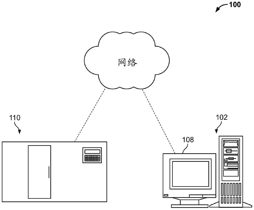
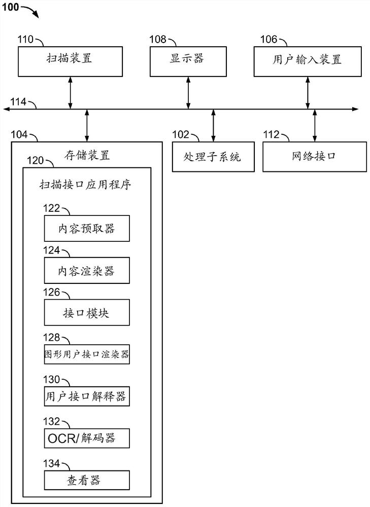
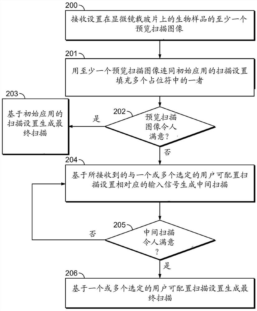

Image

  • Digital pathology scanning interface and workflow
  • Digital pathology scanning interface and workflow
  • Digital pathology scanning interface and workflow

Examples


Embodiment approach

[0120] In some embodiments, a method of utilizing a scanning device to obtain a high-resolution scan of a biological sample disposed on a substrate, the method comprising: (i) receiving, on a graphical user interface, information corresponding to user-configurable scan settings; a first user input; (ii) receiving a second user input on the graphical user interface to initiate a scan based on the series of user inputs received corresponding to said user-configurable scan settings; and (iii) said displaying a visualization of one or more placeholders on the graphical user interface, the one or more placeholders populated with one or more of scanning operation status information, image data, and at least a portion of the user-configurable scanning settings . In some embodiments, the method further includes receiving a third user input to store the image data. In some embodiments, said storing of said image data further comprises generating a high resolution scan of said image da...


PUM


Abstract

Disclosed herein is a method of acquiring a high-resolution scan of a biological sample disposed on a substrate with a scanning device, the method comprising: receiving, on a graphical user interface,a first user input corresponding to user configurable scanning settings; receiving, on a graphical user interface, a second user input to initiate scanning based on the received series of user inputscorresponding to user configurable scanning settings; and displaying, on the graphical user interface, a visualization of one or more placeholders populated with one or more of scanning operation status information, image data, and at least a portion of the user configurable scanning settings.

Description

[0001] Cross References to Related Applications [0002] This application claims the benefit of the filing date of U.S. Provisional Patent Application No. 62 / 639,375, filed March 6, 2018, the disclosure of which is hereby incorporated by reference in its entirety. Background technique [0003] Histopathology refers to the microscopic examination of various forms of biological samples. Specifically, in clinical medicine, histopathology refers to the examination of a biopsy or surgical sample by a pathologist after the sample has been processed and histological sections placed on glass specimen slides. Cytopathology is a branch of pathology that studies and diagnoses disease at the cellular level. It is often used as an aid in the diagnosis of cancer, but also in the diagnosis of certain infectious diseases and other inflammatory conditions, as well as thyroid lesions, diseases involving sterile body cavities (peritoneum, pleura, and cerebrospinal cord) and a wide range of othe...

Claims


Application Information

Patent Timeline
Patent Type & Authority Applications(China)
IPC IPC(8): G16H30/00
CPCG16H30/00G16H40/67G16H10/40G16H30/20G16H30/40G02B21/0036G02B21/006G02B21/34
Inventor M·德科斯塔D·普雷托里厄斯P·德索萨库托埃桑托斯
Owner VENTANA MEDICAL SYST INC
Learn More