Lung parenchyma CT image segmentation method based on weighted full convolutional neural network

A convolutional neural network and CT image technology, applied in the field of medical image processing, can solve problems such as incorrect segmentation, improve reliability, improve semantic segmentation performance, and reduce model redundant calculations

Pending Publication Date: 2020-11-03
BEIJING UNIV OF TECH
View PDF2 Cites 8 Cited by
  • Summary
  • Abstract
  • Description
  • Claims
  • Application Information

AI Technical Summary

Problems solved by technology

The overall classification accuracy of the lung parenchyma is high, but some small nodules around the lung wall are often not correctly segmented

Method used


Image

  • Lung parenchyma CT image segmentation method based on weighted full convolutional neural network
  • Lung parenchyma CT image segmentation method based on weighted full convolutional neural network
  • Lung parenchyma CT image segmentation method based on weighted full convolutional neural network

Examples


Embodiment Construction

[0049] The present invention can automatically process lung CT images and extract lung parenchymal regions; the present invention uses new technical means to improve the segmentation accuracy of lesion regions in lung parenchyma, and provide more reliable information support for clinical diagnosis.

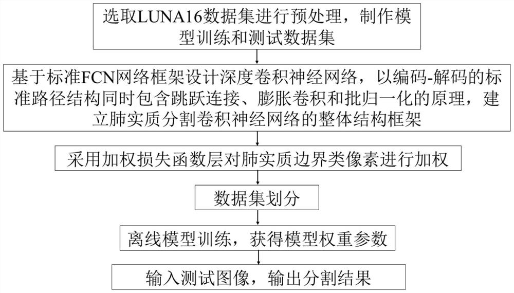
[0050] Such as process figure 1 As shown, a deep convolutional neural network lung parenchymal segmentation method based on a weighted loss function includes the following 6 steps:

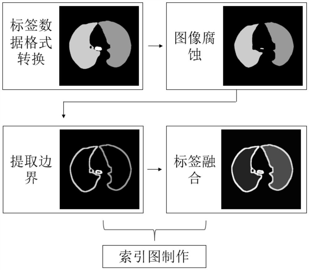
[0051] 1. Select LUNA16 (Lung Nodule Analysis 16, https: / / luna16.grand-challenge.org / ) lung CT image data set for preprocessing, and make model training and test data sets;

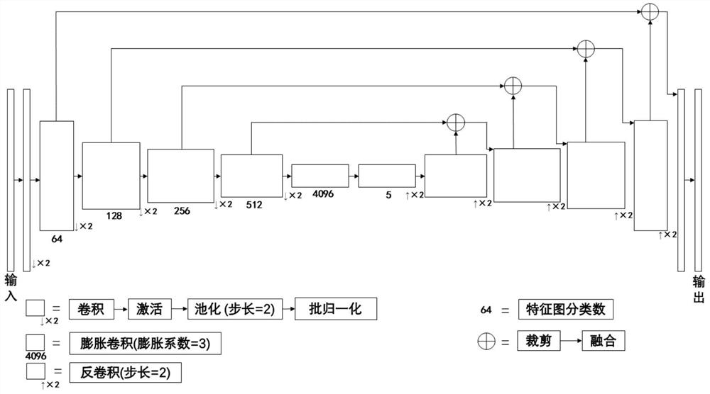
[0052] 2. Design a deep convolutional neural network based on the standard FCN network framework, and use the standard encoding-decoding path structure to include the principles of jump connection, dilation convolution and batch normalization, and establish the overall structure of the lung parenchymal segmentation convolutional neural n...


PUM


Abstract

The invention discloses a lung parenchyma CT image segmentation method based on a weighted full convolutional neural network, and belongs to the field of medical image processing. The method comprisesthe following steps: selecting a public lung data set for preprocessing, and extracting a lung parenchyma boundary in a labeled image as a semantic category; designing an improved network structure based on a standard full convolutional neural network framework, and establishing an overall structure framework of the pulmonary parenchyma segmentation convolutional neural network by using a principle that a standard path structure for encoding and decoding simultaneously comprises jump connection, expansion convolution and batch normalization; adopting a weighted loss function layer; dividing the data set; carrying out offline model training out to acquire model weight parameters; inputting a test image and outputting a segmentation result by an output layer through layer-by-layer feedforward of a network. According to an existing lung parenchyma segmentation method, a segmentation missing phenomenon is prone to occurring in a focus area in lung parenchyma, and correct segmentation of the focus area in lung parenchyma segmentation can be effectively improved through enhancement processing on important pixels.

Description

Technical field [0001] The invention relates to the field of medical image processing, in particular to a lung parenchymal CT image segmentation method based on a weighted full convolutional neural network. Background technique [0002] Relevant studies have shown that early detection and timely treatment of lung cancer can greatly improve the cure rate of lung cancer patients. CT has been proven to be an effective medical imaging technology for the diagnosis of lung diseases and is widely used in lung cancer detection and diagnosis. However, it is difficult for doctors to distinguish suspected lesion areas in a large number of CT slices with their eyes. The research and application of computer-aided diagnosis systems will help improve the accuracy and objectivity of diagnosis and reduce the workload of diagnosis. In the design of a computer-aided diagnosis system for lungs, accurate extraction of lung parenchymal regions is an important prerequisite step that affects the accurac...

Claims


Application Information

Patent Timeline
Patent Type & Authority Applications(China)
IPC IPC(8): G06T7/11G06T7/136G06T7/155G06N3/04G06N3/08
CPCG06T7/11G06T7/136G06T7/155G06N3/084G06T2207/10081G06T2207/20081G06T2207/20084G06T2207/30061G06T2207/20221G06N3/045
Inventor 林岚吴玉超吴水才
Owner BEIJING UNIV OF TECH
Why Eureka
Try Eureka