Dry immunofluorescence quantitative method heparin binding protein (HBP) detection kit

A heparin-binding protein and detection kit technology, applied in the field of in vitro diagnostic immunological assay analysis, can solve the problems of low accuracy and stability, inability to perform bedside detection, harsh storage conditions, etc. Difficulty, low cost effect of inconsistent storage conditions

Pending Publication Date: 2020-11-06
QINGDAO HIGHTOP BIOTECH
View PDF7 Cites 5 Cited by
  • Summary
  • Abstract
  • Description
  • Claims
  • Application Information

AI Technical Summary

Benefits of technology

This patented technology describes an assay that uses specific chemical agents called Hep B Phe proteins to detect certain types of molecules associated with inflammatory processes such as platelet aggregation or leukocyte activation during blood clotting. These techniques have potential applications across various fields including medical diagnosis, biological researches, environmental monitoring, etc., especially when combined together into one device.

Problems solved by technology

This patented technical problem addressed by the inventors relates to detecting biologics called liver binding proteins (LPA). These molecules bind strongly to calcium released into blood vessels during hemorrhaging caused by bleeding due to various causes like surgery or trauma. They were found to accumulate at sites where these damaged areas occur more frequently compared to normal ones. By measuring their concentrations over hours after injury, we could predict if any damage will develop quickly enough to trigger vasospasm causing fluid leaks.

Method used


Image

  • Dry immunofluorescence quantitative method heparin binding protein (HBP) detection kit
  • Dry immunofluorescence quantitative method heparin binding protein (HBP) detection kit
  • Dry immunofluorescence quantitative method heparin binding protein (HBP) detection kit

Examples


Embodiment 1

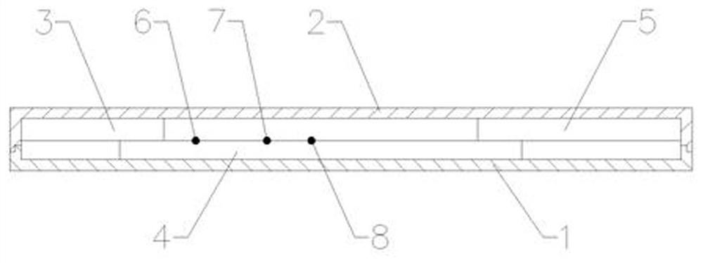
[0035] Such as figure 1 , figure 2 and image 3 As shown, a dry-type immunofluorescence quantitative method heparin-binding protein (HBP) detection kit, including a lower cover and an upper cover 2 and a reagent strip, the lower cover 1 and the upper cover 2 are snap-connected to each other, and the lower cover 1 and the upper cover 2 An accommodating cavity is formed between them, and the reagent strip is located in the accommodating cavity, and the upper cover 2 is provided with a sampling hole 9 and a detection window 10, and the reagent strip includes an absorbent paper 5, a nitrocellulose membrane 4 and a sample pad 3, and the absorbent The paper 5 is located at one end of the nitrocellulose membrane 4, the sample pad 3 is located at the other end of the nitrocellulose membrane 4, and the nitrocellulose membrane 4 is sequentially coated with a quality control C line 8, a detection T line 7, and a fluorescent The antibody solid phase line 6, the sample injection hole 9 ...

Embodiment 2

[0053] Such as figure 1 , figure 2 and image 3 As shown, a dry-type immunofluorescence quantitative method heparin-binding protein (HBP) detection kit, including a lower cover and an upper cover 2 and a reagent strip, the lower cover 1 and the upper cover 2 are snap-connected to each other, and the lower cover 1 and the upper cover 2 An accommodating cavity is formed between them, and the reagent strip is located in the accommodating cavity, and the upper cover 2 is provided with a sampling hole 9 and a detection window 10, and the reagent strip includes an absorbent paper 5, a nitrocellulose membrane 4 and a sample pad 3, and the absorbent The paper 5 is located at one end of the nitrocellulose membrane 4, the sample pad 3 is located at the other end of the nitrocellulose membrane 4, and the nitrocellulose membrane 4 is sequentially coated with a quality control C line 8, a detection T line 7, and a fluorescent The antibody solid phase line 6, the sample injection hole 9 ...

Embodiment 3

[0070] Embodiment 3: as figure 1 , figure 2 and image 3 As shown, a dry-type immunofluorescence quantitative method heparin-binding protein (HBP) detection kit, including a lower cover and an upper cover 2 and a reagent strip, the lower cover 1 and the upper cover 2 are snap-connected to each other, and the lower cover 1 and the upper cover 2 An accommodating cavity is formed between them, and the reagent strip is located in the accommodating cavity, and the upper cover 2 is provided with a sampling hole 9 and a detection window 10, and the reagent strip includes an absorbent paper 5, a nitrocellulose membrane 4 and a sample pad 3, and the absorbent The paper 5 is located at one end of the nitrocellulose membrane 4, the sample pad 3 is located at the other end of the nitrocellulose membrane 4, and the nitrocellulose membrane 4 is sequentially coated with a quality control C line 8, a detection T line 7, and a fluorescent The antibody solid phase line 6, the sample injectio...


PUM


Abstract

The invention provides a dry immunofluorescence quantitative method heparin binding protein (HBP) detection kit. The kit comprises a reagent strip, the reagent strip comprises absorbent paper, a nitrocellulose membrane and a sample pad, the absorbent paper is located at one end of the nitrocellulose membrane, the sample pad is located at the other end of the nitrocellulose membrane, and the nitrocellulose membrane is sequentially coated with a quality control C line, a detection T line and a fluorescent antibody solidus; a novel fluorescence labeling process, a sample pad treating fluid and afluorescence marker immobilization process are adopted to rapidly, accurately and quantitatively detect heparin binding protein HBP in human plasma with high sensitivity, so that the problems that labeled fluorescent substances are unstable and later process control is not facilitated are solved; meanwhile, the fluorescent antibody is immobilized on the nitrocellulose membrane instead of a specialfluorescent binding pad, so that the precision is remarkably improved on the basis of widening the storage conditions of the kit.

Description


Claims


Application Information

Patent Timeline
Owner QINGDAO HIGHTOP BIOTECH
Why Eureka
Try Eureka