Medical image processing method, device, equipment and storage medium

A technology of medical images and processing methods, applied in image data processing, image analysis, medical informatics, etc.

Active Publication Date: 2021-02-09
PING AN TECH (SHENZHEN) CO LTD
View PDF9 Cites 0 Cited by
  • Summary
  • Abstract
  • Description
  • Claims
  • Application Information

AI Technical Summary

Problems solved by technology

[0004] The main purpose of the present invention is to solve the technical problem that the existing medical image extension technology is difficult to generate new bionic abnormal tissue images based on the existing abnormal tissue images

Method used


Image

  • Medical image processing method, device, equipment and storage medium
  • Medical image processing method, device, equipment and storage medium
  • Medical image processing method, device, equipment and storage medium

Examples


Embodiment Construction

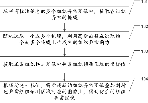
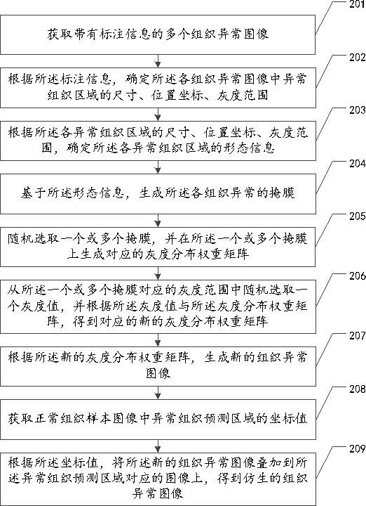
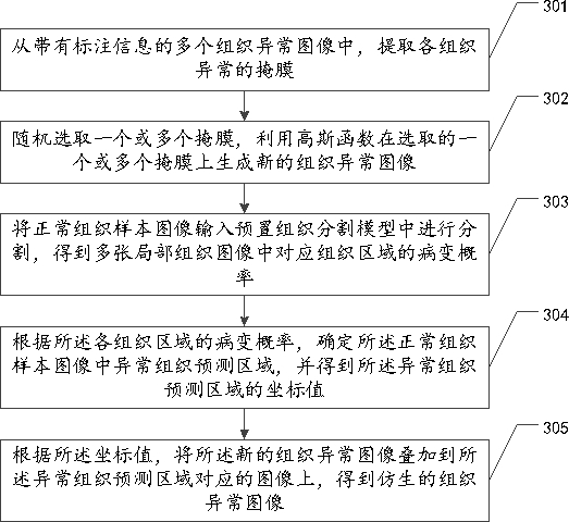
[0064] Embodiments of the present invention provide a medical image processing method, device, device, and storage medium. The method includes extracting the mask of each tissue abnormality from a plurality of abnormal tissue images with annotation information; randomly selecting one or more Masks, use Gaussian function to generate new abnormal tissue images on one or more selected masks; obtain the coordinate values ​​of the abnormal tissue prediction area in the normal tissue sample image; according to the coordinate values, the new tissue The abnormal image is superimposed on the image corresponding to the abnormal tissue prediction area to obtain a bionic abnormal tissue image. The invention realizes the bionic of the abnormal tissue image, and can control the shape, size, gray scale distribution and generation position of the lesion.

[0065] The terms "first", "second", "third", "fourth", etc. (if any) in the description and claims of the present invention and the above ...


PUM


Abstract

The present invention relates to the field of artificial intelligence, and discloses a medical image processing method, device, equipment and storage medium. The method includes: extracting the mask of each tissue abnormality from a plurality of abnormal tissue images with label information; randomly selecting One or more masks, using a Gaussian function to generate a new abnormal tissue image on the selected one or more masks; obtaining the coordinate value of the abnormal tissue prediction area in the normal tissue sample image; according to the coordinate value, the The new abnormal tissue image is superimposed on the image corresponding to the abnormal tissue prediction area to obtain a bionic abnormal tissue image. The present invention also relates to block chain technology, and the abnormal image of the tissue is stored in the block chain. The invention realizes the bionic of abnormal tissue images, and can control the lesion shape, size, gray scale distribution and generation position.

Description

technical field [0001] The present invention relates to the field of artificial intelligence, in particular to a medical image processing method, device, equipment and storage medium. Background technique [0002] With the rapid development of computing and information technology, the use of deep learning to realize intelligent diagnosis and assistance of medical images has become a current hot spot. Since deep learning technology is trained to obtain a model through the network's autonomous learning of data features, the performance of the model is closely related to the sample size used for training. The larger the sample size manually labeled, the better the model performance. Taking cerebral hemorrhage data as an example, although brain CT data are increasing at a certain rate every year, because cerebral hemorrhage is not a common disease, it is difficult to collect positive data with hemorrhage, and most of them are negative data without disease , even if a certain am...

Claims


Application Information

Patent Timeline
Patent Type & Authority Patents(China)
IPC IPC(8): G06K9/32G06K9/34G06K9/62G06T7/00G16H50/20
CPCG06T7/0012G16H50/20G06V10/25G06V10/267G06F18/2415
Inventor 陈凯星周鑫
Owner PING AN TECH (SHENZHEN) CO LTD
Why Eureka
Try Eureka