Blood leukocyte image segmentation method based on UNet++ and ResNet

An image segmentation and white blood cell technology, which is applied in the field of image processing to achieve good robustness and improve segmentation accuracy.

Pending Publication Date: 2020-12-11
MINJIANG UNIV
View PDF0 Cites 20 Cited by
  • Summary
  • Abstract
  • Description
  • Claims
  • Application Information

AI Technical Summary

Problems solved by technology

[0006] The purpose of the present invention is to improve the leukocyte segmentation accuracy in the blood leukocyte image segmentation, and provide a blood leukocyte image segmentation method based on UNet++ and ResNet, which can not only significantly improve the segmentation accuracy, but also has the advantages of different collection environments and preparation techniques for leukocyte images. good robustness

Method used


Image

  • Blood leukocyte image segmentation method based on UNet++ and ResNet
  • Blood leukocyte image segmentation method based on UNet++ and ResNet
  • Blood leukocyte image segmentation method based on UNet++ and ResNet

Examples


Embodiment Construction

[0028] The technical solution of the present invention will be specifically described below in conjunction with the accompanying drawings.

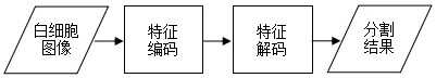
[0029] The invention provides a blood leukocyte image segmentation method based on UNet++ and ResNet, comprising:

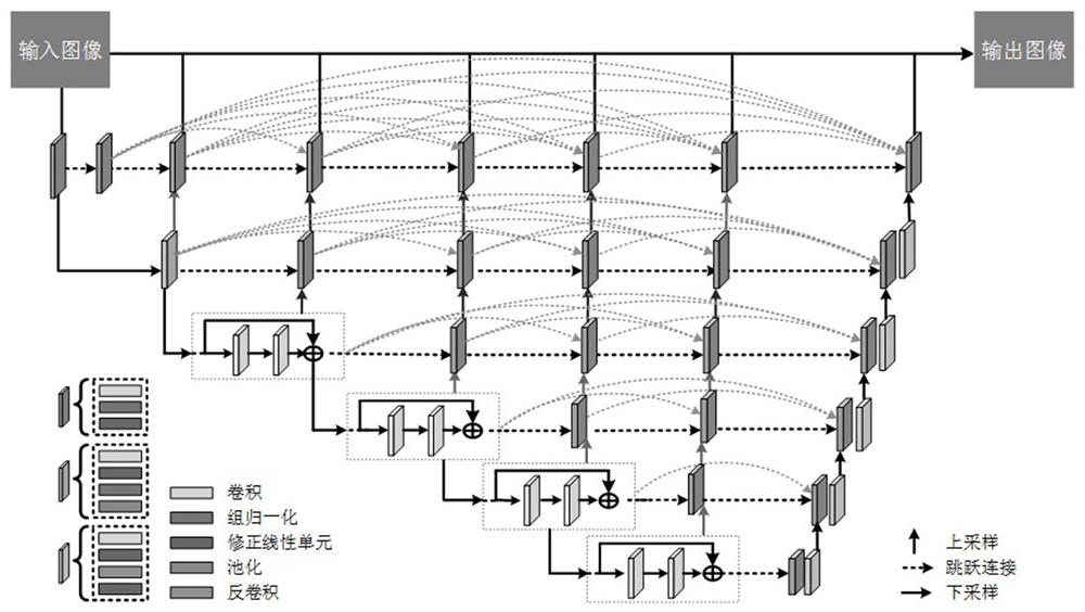
[0030] Feature encoding stage: A context-aware feature encoder with convolutional blocks and residual blocks is used to extract multi-scale feature maps, i.e. image shallow features;

[0031] Feature decoding stage: A feature decoder with convolution and deconvolution is used to resize the multi-scale feature map, that is, image deep features, to achieve end-to-end white blood cell segmentation. In the feature decoding stage, a feature decoder composed of convolution and deconvolution is used to reconstruct the segmentation mask of white blood cells, and the segmentation of white blood cells is realized through pixel-level classification.

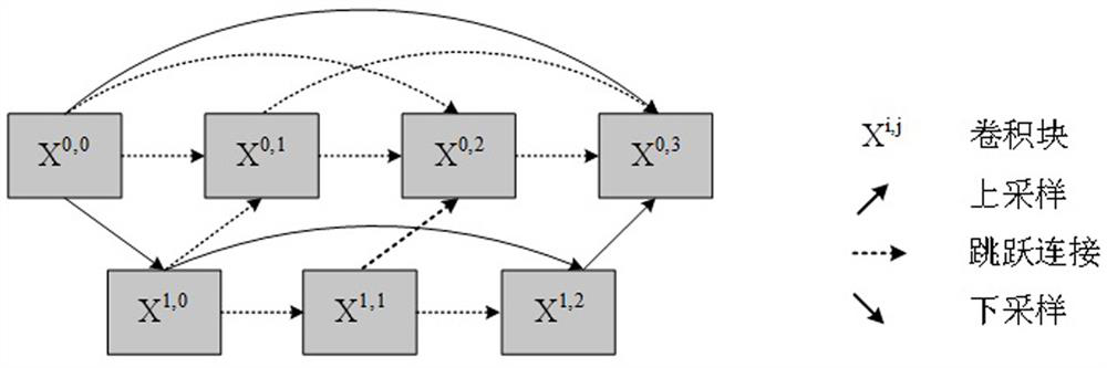
[0032] The feature decoding stage also uses a feature fusion structure of hybrid ski...


PUM


Abstract

The invention relates to a blood leukocyte image segmentation method based on UNet++ and ResNet. The method comprises the following steps: firstly, extracting multi-scale image shallow features by using an encoder with a convolution block and a residual block; extracting deep features of the image by using a decoder with convolution and deconvolution, and fusing shallow features and deep featuresby using mixed jump connection so as to reduce a semantic gap between the shallow features and the deep features; finally, designing a loss function based on the cross entropy and the Tversky index, guiding the model to learn effective image features by calculating a loss function value of each layer, and solving the problem of low training efficiency caused by imbalance of sample categories in aconventional segmentation loss function.

Description

technical field [0001] The invention belongs to the technical field of image processing, and in particular relates to a blood leukocyte image segmentation method based on UNet++ and ResNet. Background technique [0002] Information such as the total number of WBC (White Blood Cell, formerly known as Leukocyte) in the blood, the proportion and shape of various types of white blood cells are important indicators for diagnosing leukemia and other human blood diseases. An important part of the blood routine examination in the hospital is the differential count and abnormal morphology analysis of white blood cells. At present, domestic hospitals usually use a blood cell analyzer based on the electrical impedance method (physical method) plus the flow analysis method (physical-chemical method) to perform differential counting of blood cells. When the blood cell count results are abnormal or the attending doctor suspects that the patient has a blood disease, the laboratory doctor ...

Claims


Application Information

Patent Timeline
Patent Type & Authority Applications(China)
IPC IPC(8): G06T7/11G06K9/62G06N3/04G06N3/08
CPCG06T7/11G06N3/08G06T2207/30004G06N3/045G06F18/241G06F18/253
Inventor 李佐勇卢妍邵振华钟智雄樊好义
Owner MINJIANG UNIV
Why Eureka
Try Eureka