Imaging method and device for four-dimensional ultrasonic guided puncture

A three-dimensional ultrasound and ultrasound technology, applied in the field of medical testing, can solve the problems of not being able to see the needle tip of the puncture needle, not being able to reflect the spatial position relationship, and the influence of the reflected echo signal strength, etc.

Active Publication Date: 2020-12-29
深圳蓝影医学科技股份有限公司
View PDF5 Cites 2 Cited by
  • Summary
  • Abstract
  • Description
  • Claims
  • Application Information

AI Technical Summary

Problems solved by technology

[0003] However, the current ultrasound-guided puncture technology still has limitations: the image of the puncture needle and the ultrasonic sound beam of the target tissue are not on the same plane, and the phenomenon that the needle tip of the puncture needle cannot be seen occurs from time to time. High coordination ability; the echo signal strength of the puncture needle is affected by the emission angle of the ultrasonic sound beam, and the mirror reflection of the puncture needle will cause the needle tip image to be unclear, which will affect the doctor's observation
[0004] The currently proposed and adopted technologies to overcome these deficiencies largely depend on the professional background and operating skills of ultrasound doctors. The main disadvantages are: the image of the puncture needle is not on the same plane as the 2D image of the target tissue, and the puncture needle is not clear enough. Both images make it difficult for doctors to capture the position of the needle tip relative to the target tissue; although real-time 3D imaging can directly capture the information of the puncture needle in the scanning space, so far, there is still no better visualization method to distinguish the puncture needle from the target tissue. The tissue information is clearly displayed to intuitively reflect the spatial position information of the puncture needle. The common method is to display the needle tip information on multiple two-dimensional imaging sections, but it still cannot reflect the spatial position relationship
These shortcomings have brought difficulties for doctors to dynamically track the insertion trajectory of the puncture needle in real time, guide the needle body to reach the target in the shortest path, and avoid needle tip damage to important tissues and organs.

Method used


Image

  • Imaging method and device for four-dimensional ultrasonic guided puncture
  • Imaging method and device for four-dimensional ultrasonic guided puncture
  • Imaging method and device for four-dimensional ultrasonic guided puncture

Examples


Embodiment Construction

[0051] In order to make the purpose, features and advantages of the present application more obvious and understandable, the present application will be further described in detail below in conjunction with the accompanying drawings and specific implementation methods. Apparently, the described embodiments are some of the embodiments of the present application, but not all of them. Based on the embodiments in this application, all other embodiments obtained by persons of ordinary skill in the art without creative efforts fall within the protection scope of this application.

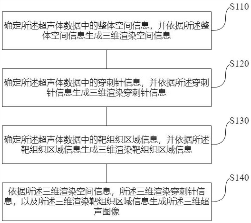
[0052] It should be noted that, in any embodiment of the present invention, a series of steps are also included before performing the method steps disclosed in the present invention: collecting the front-end data of the ultrasonic probe; performing three-dimensional reconstruction calculation on the collected front-end data processing, and performing three-dimensional post-processing on the front-end data...


PUM


Abstract

The invention provides an imaging method and device for four-dimensional ultrasonic guided puncture. The method is applied to generating a real-time three-dimensional ultrasonic image by acquiring real-time ultrasonic volume data to assist medical staff in clinical guided puncture, wherein the ultrasonic volume data is a set of data of a plurality of two-dimensional ultrasonic images which are continuous in time. The method comprises the steps of determining overall space information in the ultrasonic volume data, and generating three-dimensional rendering space information according to the overall space information; determining puncture needle information in the ultrasonic volume data, and generating three-dimensional rendering puncture needle information according to the puncture needleinformation; determining target tissue area information in the ultrasonic volume data, and generating three-dimensional rendering target tissue area information according to the target tissue area information; and generating a three-dimensional ultrasonic image according to the three-dimensional rendering space information, the three-dimensional rendering puncture needle information and the three-dimensional rendering target tissue area information. The three-dimensional ultrasonic image can clearly and visually display the spatial position of a puncture needle and the spatial anatomical structure of a target and surrounding tissue.

Description

technical field [0001] The present application relates to the field of medical detection, in particular to an imaging method and device for four-dimensional ultrasound-guided puncture. Background technique [0002] Puncture is mainly used in clinical examination and diagnosis to confirm the diagnosis, evaluate the prognosis or stage of the disease, and assist in deciding the treatment plan. Due to the differences in the structure of human tissues, the safety and effectiveness of puncture is a major difficulty for doctors. With the aid of imaging technology, the visual display of the puncture process and its surrounding tissue structure has greatly improved the accuracy, safety and efficiency of puncture, enhanced the confidence of clinicians, and reduced the damage to patients. Among them, compared with other imaging techniques (such as CT and MRI imaging), ultrasound-guided puncture has the advantages of real-time visualization, safety and radiation-free, convenient portab...

Claims


Application Information

Patent Timeline
Patent Type & Authority Applications(China)
IPC IPC(8): A61B17/34A61B8/08
CPCA61B17/3403A61B8/483A61B8/0841A61B2017/3413
Inventor 魏芳丁浩邢锐桐孙瑞超李彬陈晶
Owner 深圳蓝影医学科技股份有限公司
Why Eureka
Try Eureka