Method for real-time superposition of three-dimensional medical image and patient and operation auxiliary system

A medical imaging and 3D imaging technology, applied in surgical navigation systems, surgery, medical science, etc., can solve problems such as inaccurate matching between 3D medical images and patients' bodies, frequent switching of sight lines, etc., to reduce surgical risks, improve matching speed, The effect of high coincidence matching accuracy

Pending Publication Date: 2021-01-05
沈阳蓝软智能医疗科技有限公司 +1
View PDF0 Cites 7 Cited by
  • Summary
  • Abstract
  • Description
  • Claims
  • Application Information

AI Technical Summary

Problems solved by technology

[0003] The present invention aims to solve the problem of inaccurate matching between the three-dimensional medical image and the patient's body when the doctor compares the patient's three-dimensional medical image, locates the corresponding tissue structure of the patient's body, formulates the opera...

Method used


Image

  • Method for real-time superposition of three-dimensional medical image and patient and operation auxiliary system
  • Method for real-time superposition of three-dimensional medical image and patient and operation auxiliary system
  • Method for real-time superposition of three-dimensional medical image and patient and operation auxiliary system

Examples


Embodiment 1

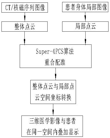
[0080] Attached below figure 1 The flow chart of the method for real-time overlapping of three-dimensional medical images and patients illustrates this embodiment 1:

[0081] A method for real-time overlapping of a three-dimensional medical image and a patient, the method being:

[0082] Convert the patient's CT sequence image or MRI sequence image into an overall point cloud, and reconstruct the 3D model of the patient's CT sequence image or MRI sequence image to form a 3D medical image;

[0083] Obtaining an image corresponding to a CT sequence image or a nuclear magnetic sequence image on the patient's body, the image is a partial image of the patient's body, and converting the partial image into a local point cloud;

[0084] The overall point cloud and the local point cloud are registered using the Super-4PCS algorithm, and the spatial coordinates of the overall point cloud and the spatial coordinates of the local point cloud are determined to accurately coincide in the s...

Embodiment 2

[0088] According to the method for real-time overlapping of three-dimensional medical images and patients described in embodiment 1, the extraction method of the whole point cloud of the present invention is specifically described,

[0089] The extraction method of the whole point cloud is:

[0090] Select an appropriate threshold in the patient's CT sequence images or MRI sequence images;

[0091] Since there is a significant difference in the pixel gray value of the target area and the background area of ​​the patient's CT image or nuclear magnetic image, so the present invention uses a threshold segmentation algorithm to segment the patient's CT image or nuclear magnetic image, and the selection process of the appropriate threshold is:

[0092] (1) Select a pre-estimated threshold T according to the pixel gray value of the target area and the background area in the patient's CT image or MRI image;

[0093] (2) Segment the CT image or MRI image of the patient with the thres...

Embodiment 3

[0102] According to the method for real-time overlapping of the three-dimensional medical image and the patient described in embodiment 1, the method for obtaining the local point cloud is:

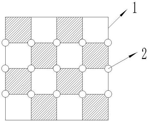
[0103] Find the characteristic region corresponding to the CT sequence image or nuclear magnetic sequence image obtained before applying the method on the patient's body, use a depth image acquisition device to obtain continuous depth images of the characteristic region, and convert the continuous depth of the characteristic region Images are converted into local point clouds using threshold segmentation.


PUM


Abstract

The invention relates to a method for real-time superposition of a three-dimensional medical image and a patient and an operation auxiliary system. The method and the operation auxiliary system are used for assisting planning and implementation of a clinical operation. The method comprises the steps that a CT sequence image or a nuclear magnetic sequence image of a patient is converted into an integral point cloud, an image of a part corresponding to the CT sequence image or the nuclear magnetic sequence image is acquired from the body of the patient, the image is a local image of the body ofthe patient, and the local image is converted into a local point cloud; the overall point cloud and the local point cloud are registered by adopting a Super-4PCS algorithm, and an optimal conversion matrix in which the space coordinates of the overall point cloud and the space coordinates of the local point cloud accurately coincide under the same space coordinate system is determined; a space coordinate of the three-dimensional medical image when the space coordinate of the three-dimensional medical image and the space coordinate of the local point cloud are accurately superposed under the same space coordinate system is determined through the optimal conversion matrix; and the three-dimensional medical image and the space coordinates thereof are transmitted to a three-dimensional image display device. According to the method, the problem of inaccurate matching between the three-dimensional medical image and the body of the patient in the existing operation process can be solved.

Description

technical field [0001] The invention relates to a method for real-time overlapping of three-dimensional medical images and patients and an operation assistance system, which are used to assist the planning and implementation of clinical operations, especially the implementation of open operations. Background technique [0002] 3D medical imaging is becoming more and more common in assisting clinical operations. It is a 3D model file generated after 3D reconstruction based on the patient's CT or MRI sequence images. Most hospitals use 3D medical imaging to display 3D medical images on the On the two-dimensional display device, the doctor locates the corresponding tissue structure of the patient's body or formulates a surgical implementation plan and plans the surgical path by comparing the patient's three-dimensional medical image. Switch the viewing angle, and then rely on the short-term memory of the 3D medical image to find the corresponding tissue structure on the patient...

Claims


Application Information

Patent Timeline
IPC IPC(8): A61B34/10A61B34/20A61B90/00
CPCA61B34/10A61B34/20A61B90/37A61B2034/101A61B2034/107A61B2034/2065A61B2090/374A61B2090/3762
Inventor 杨云鹏谢锦华孙野
Owner 沈阳蓝软智能医疗科技有限公司
Why Eureka
Try Eureka