Improved glioma segmentation method adopting cross-sequence nuclear magnetic resonance image generation

A technology of nuclear magnetic resonance images, glioma, applied in the medical field, to achieve the effect of avoiding subjective bias

Inactive Publication Date: 2021-01-08
RES & DEV INST OF NORTHWESTERN POLYTECHNICAL UNIV IN SHENZHEN +1
View PDF7 Cites 2 Cited by
  • Summary
  • Abstract
  • Description
  • Claims
  • Application Information

AI Technical Summary

Problems solved by technology

Relying on doctors to make a diagnosis will inevitably bring in the doctor's subjective bias

Method used


Image

  • Improved glioma segmentation method adopting cross-sequence nuclear magnetic resonance image generation
  • Improved glioma segmentation method adopting cross-sequence nuclear magnetic resonance image generation
  • Improved glioma segmentation method adopting cross-sequence nuclear magnetic resonance image generation

Examples


Embodiment Construction

[0026] The present invention will be further described below in conjunction with the accompanying drawings and embodiments.

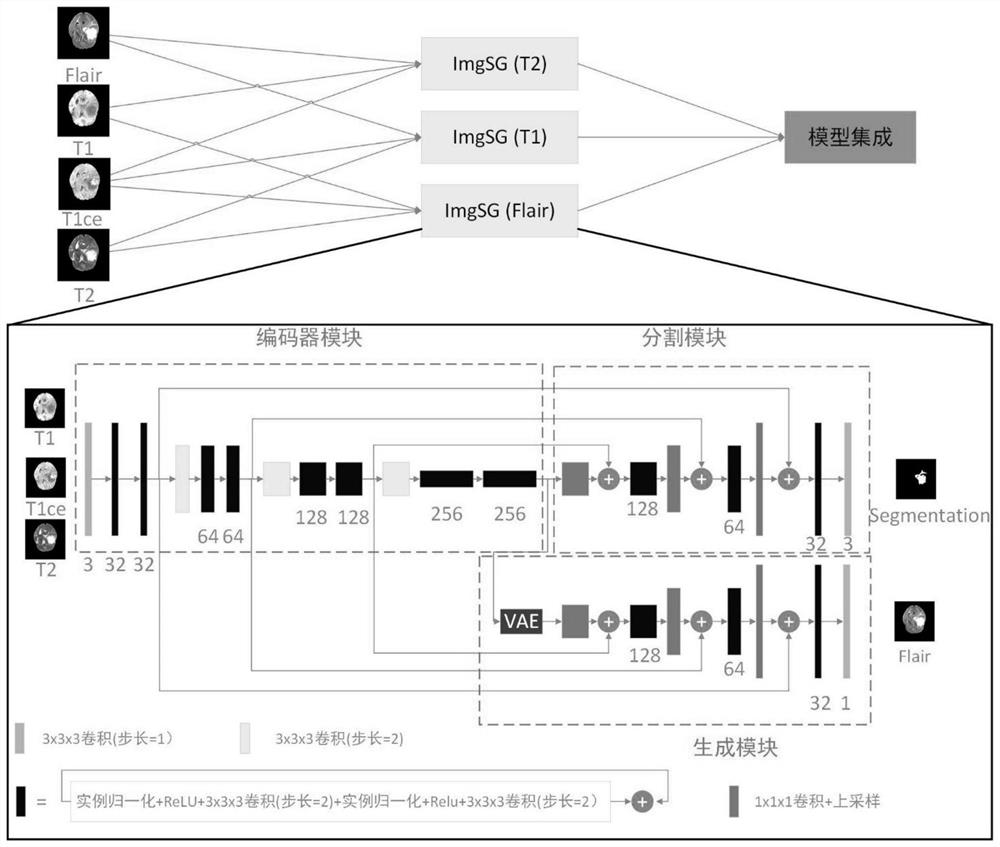
[0027] Such as figure 1 As shown, the present invention provides a method for improving glioma segmentation using cross-sequence nuclear magnetic resonance image generation, comprising the following steps:

[0028] Step 1: Due to the wide voxel range of the nuclear magnetic resonance image, the glioma nuclear magnetic resonance image is processed according to formula (1), and the voxel value of the glioma nuclear magnetic resonance image is limited within the range of [0,1];

[0029]

[0030] In the formula, x is the result of limiting the voxel value of the glioma MRI image to the range [0,1], x o is the original voxel value of glioma MRI image, x mean is the mean value of the non-zero voxel area in the glioma MRI image, and σ is the standard deviation of the non-zero voxel area in the glioma MRI image;

[0031] Step 2: Perform skull removal and ...


PUM


Abstract

The invention provides an improved glioma segmentation method adopting cross-sequence nuclear magnetic resonance image generation. According to the method, an ImgSG model composed of an encoder module, a segmentation module and a generator module is constructed; in an ImgSG model training stage, three of four sequences of a glioma nuclear magnetic resonance image are selected as input of a networkto perform glioma segmentation, the remaining sequence is used as a generation sequence to assist model segmentation, and three segmentation models with the same structure and different parameters are trained according to different generation sequences; In a test stage, a to-be-tested glioma nuclear magnetic resonance image is input into the three segmentation models, an average value of the obtained three segmentation confidence probability results is calculated, and a final glioma segmentation result is acquired by post-processing. According to the method, the current situation that a doctor judges whether glioma exists or not by reading nuclear magnetic resonance images frame by frame can be changed, and subjective deviation caused by doctor experience is avoided.

Description

technical field [0001] The invention belongs to the field of medicine, and in particular relates to a segmentation method. Background technique [0002] Glioma is the most common primary tumor in the central nervous system, accounting for more than 40% of brain tumors. In the traditional medical field, the best way to characterize a brain tumor is a biopsy. However, due to the precision of the human brain, it may cause some unpredictable consequences. Therefore, non-invasive methods represented by MRI images have become the main means of brain tumor analysis. However, the diagnosis of glioma based on MRI images is realized by doctors reading frame by frame to judge the layers. Relying on doctors for diagnosis will inevitably bring in the doctor's subjective bias. In addition, the growth rate of radiologists in my country is far behind the growth rate of medical imaging data, and there is an urgent need for automatic segmentation of glioma. Contents of the invention ...

Claims


Application Information

Patent Timeline
Patent Type & Authority Applications(China)
IPC IPC(8): G06T7/10G06T7/30G06N3/04
CPCG06T7/10G06T7/30G06T2207/20081G06T2207/30096G06T2207/30016G06N3/045
Inventor 夏勇赵国靖张建鹏
Owner RES & DEV INST OF NORTHWESTERN POLYTECHNICAL UNIV IN SHENZHEN
Why Eureka
Try Eureka