
Method and related product for processing lung lesion area images

A lesion area and lung technology, applied in the field of image processing, can solve problems such as lack of information and inability to reflect the essential attributes of the lesion area, and achieve the effect of effective human intervention

Active Publication Date: 2021-02-19
BEIJING FRIENDSHIP HOSPITAL CAPITAL MEDICAL UNIV +2
View PDF4 Cites 2 Cited by
  • Summary
  • Abstract
  • Description
  • Claims
  • Application Information

AI Technical Summary

Problems solved by technology

However, the features extracted by traditional methods contain less information, which cannot reflect the essential properties of the lesion area.

Method used


Image

  • Method and related product for processing lung lesion area images
  • Method and related product for processing lung lesion area images
  • Method and related product for processing lung lesion area images

Examples


Embodiment Construction

[0022] The technical solutions in the embodiments of the present disclosure will be clearly and completely described below in conjunction with the accompanying drawings. It should be understood that the embodiments described in this specification are only some of the embodiments provided by the present disclosure to facilitate a clear understanding of the solutions and comply with legal requirements, but not all embodiments of the present invention can be implemented. Based on the embodiments disclosed in this specification, all other embodiments obtained by those skilled in the art without making creative efforts belong to the protection scope of the present disclosure.

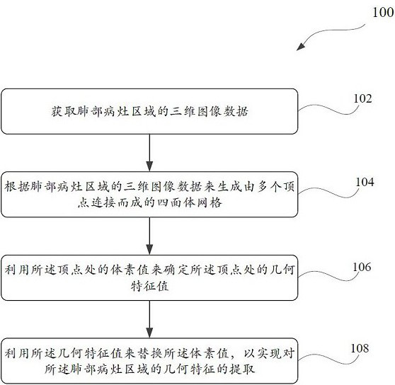
[0023] figure 1 is a flow chart illustrating a method 100 for processing images of lung lesion regions according to multiple embodiments of the present invention. It should be noted that the method 100 of the present invention can be implemented by various types of computing equipment including, for example...


PUM


Abstract

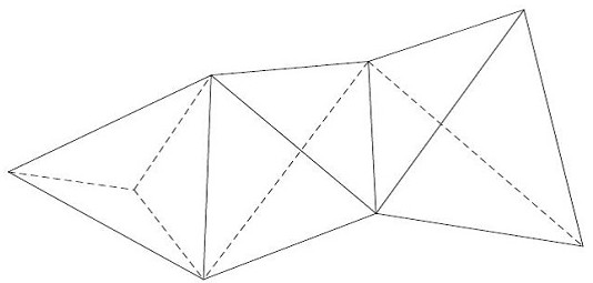
The invention discloses a method for processing a lung lesion area image and a related product. The method comprises the following steps: acquiring three-dimensional image data of a lung lesion area,generating a tetrahedral mesh formed by connecting a plurality of vertexes according to the three-dimensional image data of the lung lesion area, determining a geometric characteristic value at the vertex by utilizing the voxel value at the vertex, and replacing the voxel value with the geometric feature value to realize extraction of the geometric features of the lung lesion area. According to themethod, the high-order geometrical characteristics are extracted from the lung lesion area image, so that richer characteristic information is contained, the intrinsic geometrical attributes of the lesion area image can be reflected, and subsequent further research is facilitated.

Description

technical field [0001] The present invention relates generally to the field of image processing. More specifically, the present invention relates to a method, device and computer-readable storage medium for processing images of lung lesion areas. Background technique [0002] The image of the lesion area, especially the lung lesion area contains information that is helpful for clinical diagnosis, so the extraction of image features of the lesion area is particularly important. The current traditional processing method is to extract the radiomics features of the lesion area, and use the radiomics features for subsequent analysis and research in order to evaluate the lesion area. However, the features extracted by traditional methods contain less information, which cannot reflect the essential attributes of the lesion area. Therefore, how to extract the high-order geometric features of the lesion area and use it for lesion area analysis has become an urgent problem to be sol...

Claims


Application Information

Patent Timeline
IPC IPC(8): G06T7/00G06T17/20G06K9/32G06K9/34
CPCG06T7/0012G06T17/20G06T2207/30061G06V10/25G06V10/267
Inventor 王振常雷娜侯代伦李维任玉雪吕晗魏璇张茗昱陈伟吴伯阳
Owner BEIJING FRIENDSHIP HOSPITAL CAPITAL MEDICAL UNIV
Learn More