

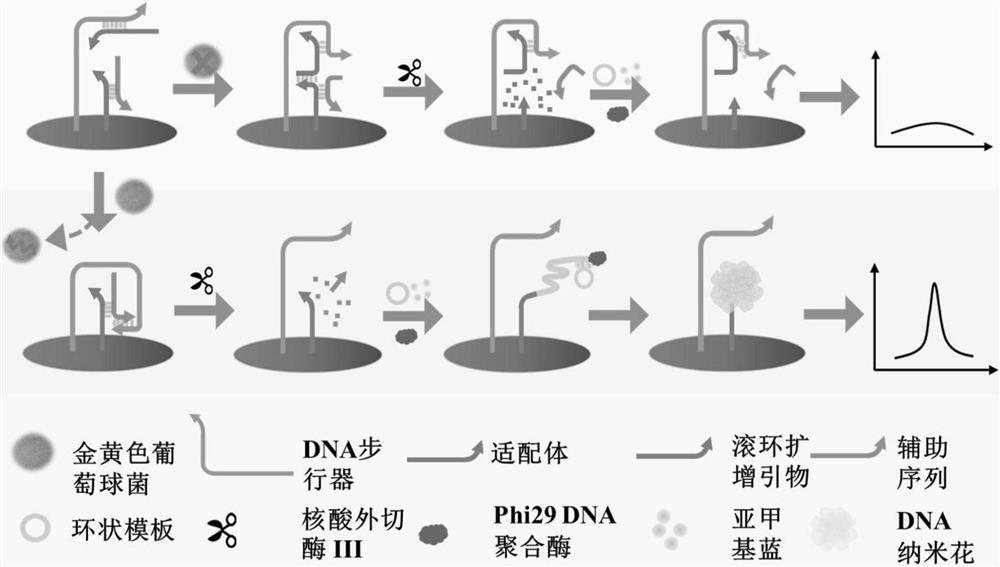
Pathogenic bacteria electrochemical detection method based on DNA walker and nanoflower structure
A detection method and pathogenic bacteria technology, applied in the field of detection, can solve the problem of fewer DNA nanoflowers, achieve the effect of increasing the effective area, broadening the detection range, and improving the detection sensitivity
- Summary
- Abstract
- Description
- Claims
- Application Information
AI Technical Summary
Benefits of technology
Problems solved by technology
Method used
Image

Examples
Embodiment 1
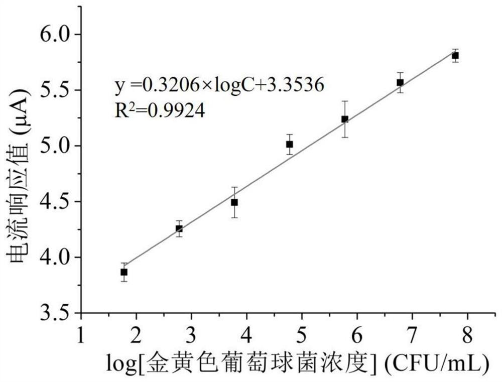
[0039] Embodiment 1: the drafting of staphylococcus aureus concentration standard curve
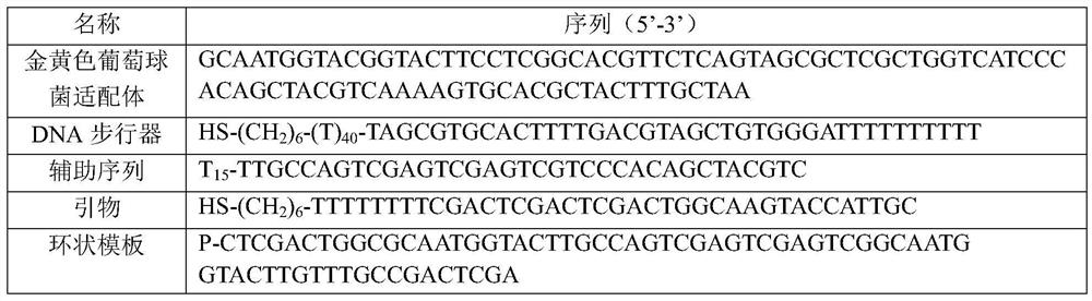
[0040] with 30% H 2 o 2 and H 2 SO 4 Prepare the piranha solution, soak the gold electrode in the piranha solution for 15 minutes to remove surface impurities, clean the electrode surface and transfer it to ultrapure water for 5 minutes of ultrasonication, and then the electrode is cleaned with 0.5mol L -1 h 2 SO 4 Perform electrochemical cleaning and electrode activation, clean the electrode again with ultrapure water, and dry it with nitrogen. 0.2μmol·L -1 DNA walker sequence and 0.4 μmol L -1 Add the aptamer to PBS buffer, 1 μmol L -1 Rolling circle amplification primers and 2 μmol L -1 The auxiliary sequence was added to PBS buffer, and the two mixtures were heat denatured at 95°C for 10 minutes, and then placed in an ice bath at 4°C for 10 minutes to form aptamer / DNA walker double strands and auxiliary sequence / rolling circle amplification primer pairs respectively. chain. ...
Embodiment 2
[0046] Embodiment 2: the mensuration of Staphylococcus aureus content in actual sample
[0047] In order to further verify the accuracy of this method in the determination of the content of Staphylococcus aureus in actual samples, Taihu Lake water and tap water without pretreatment and honey water diluted 5 times with PBS buffer were selected for the addition of Staphylococcus aureus Determination.
[0048] with 30% H 2 o 2 and H 2 SO 4 Prepare the piranha solution, soak the gold electrode in the piranha solution for 15 minutes to remove surface impurities, clean the electrode surface and transfer it to ultrapure water for 5 minutes of ultrasonication, and then the electrode is cleaned with 0.5mol L -1 h 2 SO 4 Perform electrochemical cleaning and electrode activation, clean the electrode again with ultrapure water, and dry it with nitrogen. 0.2μmol·L -1 DNA walker sequence and 0.4 μmol L -1 Add the aptamer to PBS buffer, 1 μmol L -1 Rolling circle amplification prim...
PUM
 Abstract
Description
Claims
Application Information