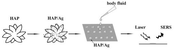
Body fluid Raman spectrum detection method of HAP/Ag composite SERS enhanced substrate
A Raman spectroscopic detection and surface-enhanced Raman technology is applied in the field of body fluid Raman spectroscopic detection of HAP/Ag composite SERS-enhanced substrates, which can solve the problems of impracticality, high laser power, sample damage, etc., and achieves short time consumption. , the effect of simple operation
- Summary
- Abstract
- Description
- Claims
- Application Information
AI Technical Summary
Problems solved by technology
Method used
Image

Examples
Embodiment 1
[0031] Preparation of Hydroxyapatite HAP
[0032] Weigh 1-1.5 g Ca(NO 3 ) 2 • 4H 2 Dissolve O in 25-50 mL of ultrapure water, stir for 10-30 min; weigh 1-1.3 g of Na 2 HPO 4 •12H 2 O was dissolved in 25-50 mL of ultrapure water; Na 2 HPO 4 •12H 2 O was added dropwise to Ca(NO 3 ) 2 • 4H 2 In the O solution, during the dropwise addition, the solution gradually changed from clear to cloudy, then the pH value of the solution was titrated with dilute nitric acid to 2.5, and the solution became clear; then 5-10 mg of cyclohexane hexacarboxylic acid was added to the solution h 6 L and 1-3g CO(NH 2 ) 2 Stir continuously for 20-50 min; carry out hydrothermal reaction of the above clear solution at 100-150°C, react for 3-5 h, and cool to room temperature naturally after the reaction; collect the product by centrifugation, wash with ethanol and ultrapure water, and finally dry in vacuum Dry in the box for 64-72 h, and then grind into hydroxyapatite nanoparticle powder.
Embodiment 2
[0034] Preparation of HAP / Ag
[0035] Weigh 3-7 mg of dried HAP powder and add it to ethanol solution containing 15 mL, 20 mM silver nitrate, and ultrasonically disperse it for 25-50 min; Add 15-30 µL of n-butylamine to it, and stir it magnetically for 30-50 min; after the reaction, the reaction product is centrifuged, washed twice with water and twice with absolute ethanol, and finally vacuum-dried and placed in The HAP / Ag nanoparticles are obtained; the prepared HAP / Ag is coated on an aluminum sheet as a SERS-enhanced substrate.
Embodiment 3
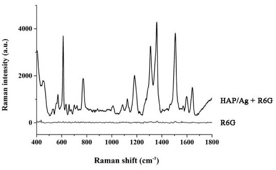
[0037] Adding R6G based on the HAP / Ag substrate proves that it has the effect of enhancing the substrate
[0038] First prepare a concentration of 10 -4 M's R6G solution. Directly take 4 µL of R6G solution and place it on an aluminum sheet with a purity of 99.99%, and dry it at room temperature for spectral detection, as shown in figure 2 As shown, there is no SERS characteristic peak in pure R6G. Then weigh 2-6 mg of HAP / Ag powder and add it to a 1.5 mL polypropylene centrifuge tube, add 60-120 μL distilled water to the centrifuge tube, ultrasonically disperse for 10-30 min, and smear the dispersed HAP / Ag on On an aluminum sheet with a purity of 99.99%, 30 µL of the R6G solution was dropped onto the HAP / Ag substrate, dried at room temperature, and detected by SERS spectrum. The laser excitation wavelength used in the detection is 785 nm, and the spectral range is 400-1800 cm -1 , the power is 10 mW, the integration time is 10 s, and the integration times are 2 times, the...
PUM
 Abstract
Description
Claims
Application Information
 - R&D Engineer
- R&D Manager
- IP Professional
- Industry Leading Data Capabilities
- Powerful AI technology
- Patent DNA Extraction
Browse by: Latest US Patents, China's latest patents, Technical Efficacy Thesaurus, Application Domain, Technology Topic.
© 2024 PatSnap. All rights reserved.Legal|Privacy policy|Modern Slavery Act Transparency Statement|Sitemap