Lung lobe segmentation system based on three-dimensional convolutional neural network
A three-dimensional convolution and neural network technology, applied in the field of image processing, can solve the problems of not meeting the daily work needs of doctors and low execution efficiency
- Summary
- Abstract
- Description
- Claims
- Application Information
AI Technical Summary
Problems solved by technology
Method used
Image

Examples
Embodiment 1
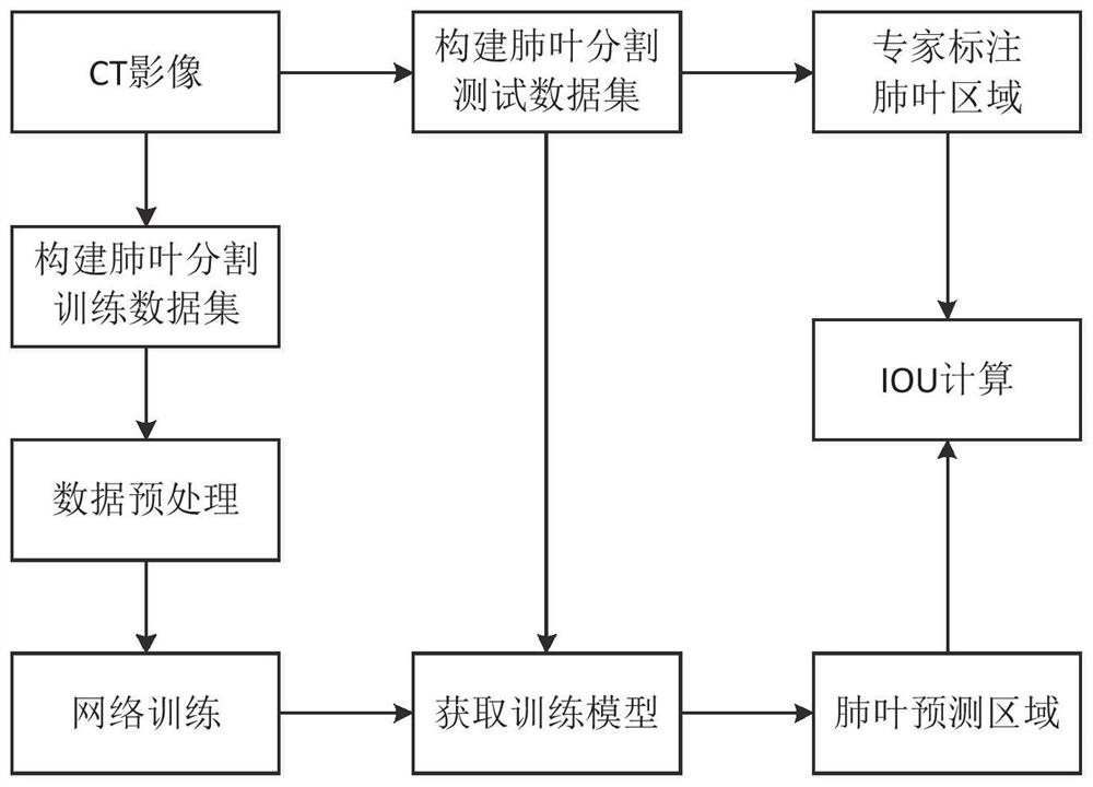
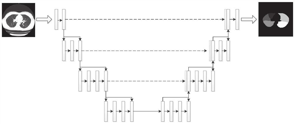
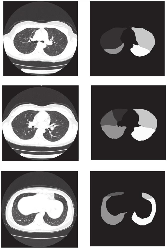
[0046] Such as figure 1 , figure 2 with image 3 As shown, Embodiment 1 of the present disclosure provides a lung lobe segmentation system based on a three-dimensional convolutional neural network, including steps:
[0047] The data acquisition module is configured to: acquire chest CT image data to be identified, and perform preprocessing;
[0048] The lung lobe segmentation module is configured to: input the preprocessed lung CT image data into a preset three-dimensional convolutional neural network model to obtain a lung lobe segmentation result.
[0049] Specifically, include the following:
[0050] S1: Select the appropriate chest CT data, and expert doctors will carry out the data labeling work, mainly through the 3DSlicer software, the 5 lung lobes of the lungs are drawn along the edges with different colors, including: left upper lobe, The lower lobe of the left lung, the upper lobe of the right lung, the middle lobe of the right lung, and the lower lobe of the ri...
Embodiment 2
[0067] Embodiment 2 of the present disclosure provides a computer-readable storage medium on which a program is stored, and when the program is executed by a processor, the following steps are implemented:
[0068] Obtain chest CT image data to be identified and perform preprocessing;
[0069] Input the preprocessed lung CT image data into the preset three-dimensional convolutional neural network model to obtain the lung lobe segmentation result;
[0070] Among them, the three-dimensional convolutional neural network model includes three down-sampling layers and three up-sampling layers, the down-sampling layer is realized by the convolution layer, and the up-sampling layer is realized by the deconvolution layer.
[0071] The detailed steps are the same as the method provided in Embodiment 1, and will not be repeated here.
Embodiment 3
[0073] Embodiment 3 of the present disclosure provides an electronic device, including a memory, a processor, and a program stored in the memory and operable on the processor, and the processor implements the following steps when executing the program:
[0074] Obtain chest CT image data to be identified and perform preprocessing;
[0075] Input the preprocessed lung CT image data into the preset three-dimensional convolutional neural network model to obtain the lung lobe segmentation result;
[0076] Among them, the three-dimensional convolutional neural network model includes three down-sampling layers and three up-sampling layers, the down-sampling layer is realized by the convolution layer, and the up-sampling layer is realized by the deconvolution layer.
[0077] The detailed steps are the same as the method provided in Embodiment 1, and will not be repeated here.
[0078] Those skilled in the art should understand that the embodiments of the present disclosure may be pr...
PUM
 Abstract
Description
Claims
Application Information
 - R&D
- Intellectual Property
- Life Sciences
- Materials
- Tech Scout
- Unparalleled Data Quality
- Higher Quality Content
- 60% Fewer Hallucinations
Browse by: Latest US Patents, China's latest patents, Technical Efficacy Thesaurus, Application Domain, Technology Topic, Popular Technical Reports.
© 2025 PatSnap. All rights reserved.Legal|Privacy policy|Modern Slavery Act Transparency Statement|Sitemap|About US| Contact US: help@patsnap.com