
Follow-up visit method and device based on medical images and computer readable storage medium

A medical imaging and imaging technology, applied in the field of medical image processing and deep learning, can solve problems such as poor work efficiency and incomplete matching between multi-phase data.

Pending Publication Date: 2021-04-20
讯飞医疗科技股份有限公司
View PDF0 Cites 3 Cited by
  • Summary
  • Abstract
  • Description
  • Claims
  • Application Information

AI Technical Summary

Problems solved by technology

[0003] Affected by factors such as the shooting environment, the multi-period data of the same user are not completely matched, and the existing multi-period follow-up method can only display the images of the target tissue in the phase of the inspection or Text, resulting in poor work efficiency

Method used


Image

  • Follow-up visit method and device based on medical images and computer readable storage medium
  • Follow-up visit method and device based on medical images and computer readable storage medium
  • Follow-up visit method and device based on medical images and computer readable storage medium

Examples


Embodiment Construction

[0023] The following will clearly and completely describe the technical solutions in the embodiments of the application with reference to the drawings in the embodiments of the application. Apparently, the described embodiments are only some of the embodiments of the application, not all of them. Based on the embodiments in this application, all other embodiments obtained by persons of ordinary skill in the art without making creative efforts belong to the scope of protection of this application.

[0024] Terms used in the embodiments of the present application are only for the purpose of describing specific embodiments, and are not intended to limit the present application. The singular forms "a", "said" and "the" used in the embodiments of this application and the appended claims are also intended to include plural forms, unless the above clearly indicates otherwise, "multiple "Generally includes at least two, but does not exclude the inclusion of at least one.

[0025] It ...


PUM


Abstract

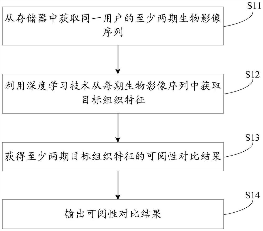
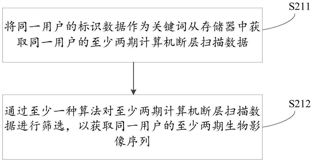
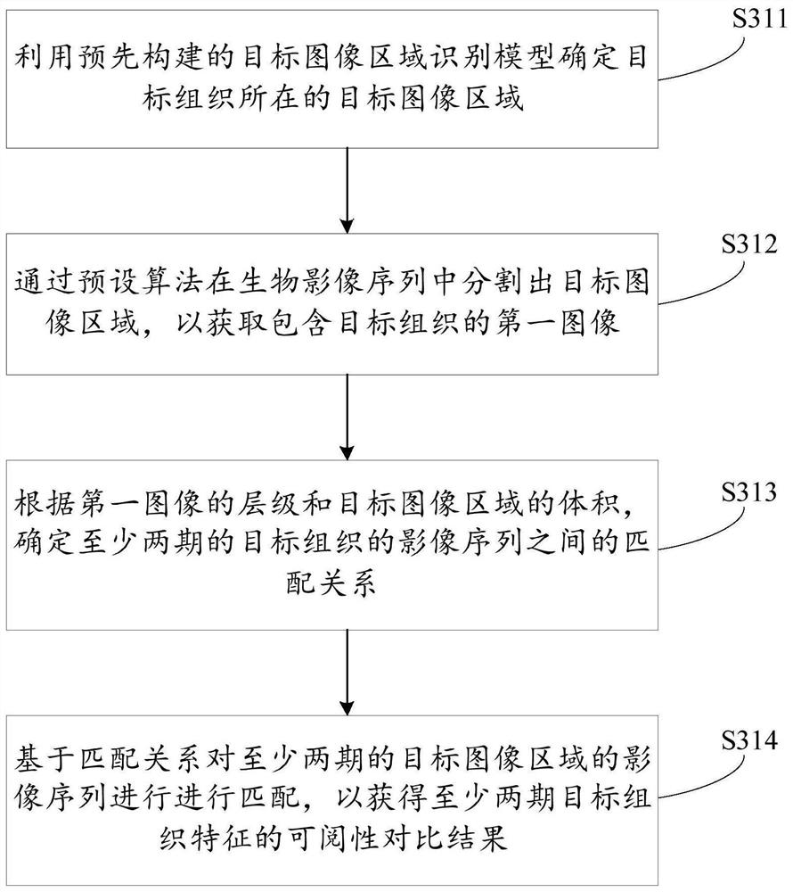
The invention discloses a follow-up visit method and device based on medical images and a computer readable storage medium. The follow-up visit method comprises the following steps: acquiring at least two stages of biological image sequences of the same user from a memory; obtaining target tissue features from each stage of biological image sequence by using a deep learning technology; obtaining a readability comparison result of at least two stages of target tissue characteristics; and outputting a readability comparison result. Through the follow-up visit method, the examination data and / or the detection conclusion of the target organization in the multi-stage examination can be visually displayed, browsing and comparison are facilitated, and the working efficiency is further improved.

Description

technical field [0001] The present application relates to the field of medical image processing and deep learning, in particular to a follow-up method, device and computer-readable storage medium based on medical images. Background technique [0002] Computed Tomography (CT) is an important imaging method for obtaining molecular information of biological tissues. With the development of medical technology, CT is often used in physical examination now, using X-ray beams, γ-rays, ultrasonic waves, etc., together with highly sensitive detectors to perform continuous cross-sectional scans around a certain part of the human body, with scan time Fast, clear image and so on. [0003] Affected by factors such as the shooting environment, the multi-period data of the same user are not completely matched, and the existing multi-period follow-up method can only display the images of the target tissue in the phase of the inspection or Text, resulting in poor work efficiency. Content...

Claims


Application Information

Patent Timeline
IPC IPC(8): G06T7/00G06T7/136G06T7/155G06T7/187G06T7/33G06T7/62G16H30/20G06N3/08
Inventor 尹孝映周璟瑜鹿晓亮葛磊
Owner 讯飞医疗科技股份有限公司
Learn More