Radiography head fixing device for cardiovascular and cerebrovascular department

A cardiovascular and cerebrovascular and fixation device technology, applied in application, medical science, patient positioning for diagnosis, etc., can solve the problem of increasing the difficulty for doctors to clearly observe the disease, unfavorable treatment of patients, and dislocation and tailing of angiographic images and other problems, to achieve the effect of reducing the support height and improving the use efficiency

Inactive Publication Date: 2021-04-23
刘燕清
View PDF0 Cites 0 Cited by
  • Summary
  • Abstract
  • Description
  • Claims
  • Application Information

AI Technical Summary

Problems solved by technology

[0002] Cardiovascular angiography is widely used in clinical practice due to its technical characteristics of small trauma, fast detection and relatively obvious results. At present, when treating cerebrovascular patients, it is necessary to perform contrast irradiation on the head of the patient, and the patient needs to lie on the Then fix the head on the radiography bed to prevent the blurred image caused by the shaking of the head and make the doctor misdiagnose. The existing radiography head fixation device for cardiovascular and cerebrovascular departments is used in radiography, because the shape of the back of the human body is different. There will be a gap between the receiving surface at the bottom and the back of the head, so that when the patient is dizzy due to the rotation of the contrast device, the patient subconsciously raises the head to drive the neck, providing arch space, resulting in dislocation and tailing of the contrast image. Increase the difficulty for doctors to clearly observe the diseased area, which is not conducive to the treatment of patients

Method used


Image

  • Radiography head fixing device for cardiovascular and cerebrovascular department
  • Radiography head fixing device for cardiovascular and cerebrovascular department
  • Radiography head fixing device for cardiovascular and cerebrovascular department

Examples


Embodiment 1

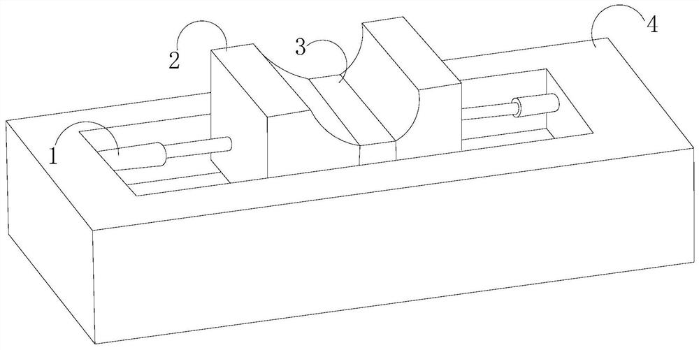
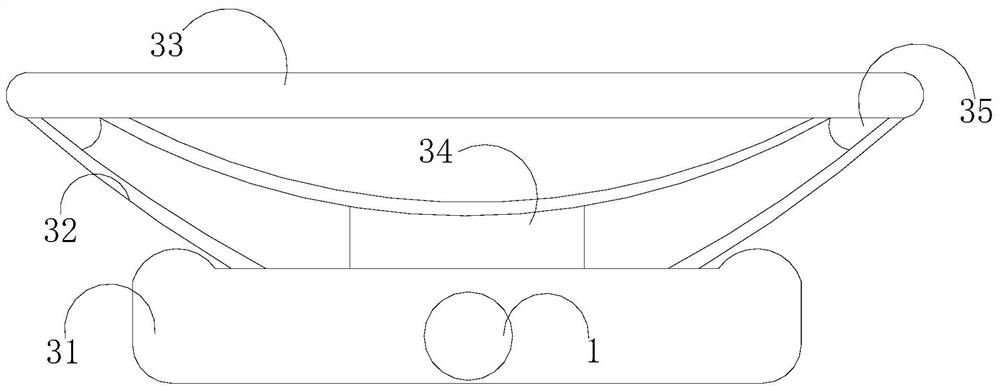
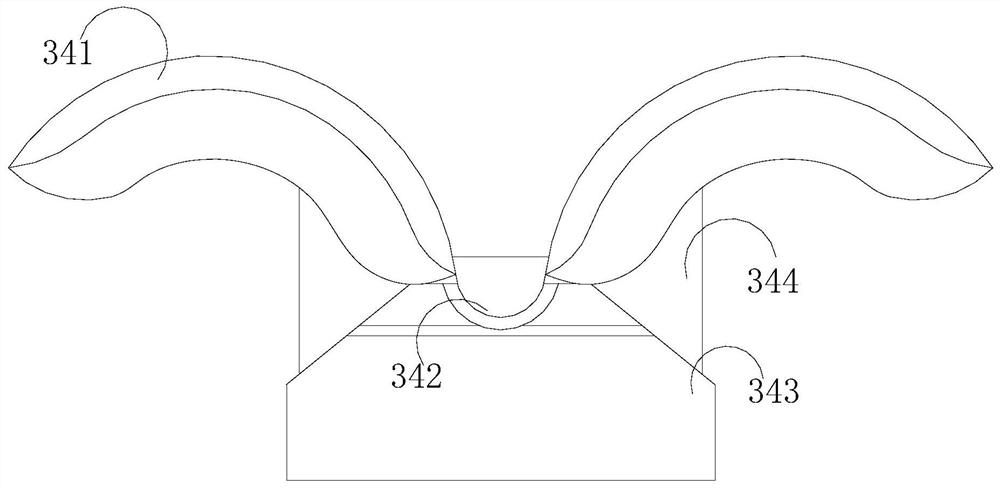
[0028] Such as Figure 1-Figure 3 As shown, the present invention provides a radiography head fixation device for cardiovascular and cerebrovascular departments. Its structure includes a telescopic rod 1, a fixed plate 2, a pressure frame 3, and a positioning frame 4. Rod 1, the top of the telescopic rod 1 is inserted on both sides of the fixed plate 2, and the middle part of the fixed plate 2 is equipped with a pressure frame 3, and the pressure frame 3 includes a base 31, a limit frame 32, and a rubber pad 33 , booster device 34, control angle 35, the base 31 is welded and connected between the fixed plates 2 through the telescopic rod 1, the limit frame 32 is embedded and connected inside the base 31, and the top end is transitionally fitted with the control angle 35 Inside the rubber pad 33, a booster device 34 is provided in the middle of the rubber pad 33. The booster device 34 includes a filling assembly 341, a balance piece 342, a socket 343, and a bearing column 344. ...

Embodiment 2

[0030] Such as Figure 4-Figure 7 As shown, on the basis of Embodiment 1, the present invention combines the mutual cooperation of the following structural components. The filling assembly 341 includes a top pad 411, a sliding guide plate 412, a partition 413, a ground block 414, and a fitting side plate 415. , the top pad 411 is fixedly connected with a fitting side plate 415 around it, and the fitting side plate 415 is gap-fitted on the bottom of the rubber pad 33, and the sliding guide plate 412 is an arc structure, and is welded and connected between the top pad 411 and the top pad 411. Between the partitions 413, the two sides of the partitions 413 are provided with ground contact blocks 414, and the balance member 342 includes a running-in ring 421, a rebound ball 422, a measuring groove 423, a protrusion 424, and a ejector device 425. The measuring groove 423 is equipped with an ejector device 425 and a bouncing ball 422, and a running-in ring 421 is fixedly connected a...


PUM


Abstract

The invention discloses a radiography head fixing device for the cardiovascular and cerebrovascular department. The radiography head fixing device structurally comprises a telescopic rod, a fixed plate, a pressure bearing frame and a positioning frame, wherein the inner side of the positioning frame is fixedly connected with the telescopic rod; the top end of the telescopic rod is inserted and embedded into the two sides of the fixed plate; the pressure bearing frame is installed in the middle of the fixed plate; and a filling assembly and a balancing part are arranged in a power assisting device. The filling assembly and the balancing part are matched in a bearing column; when a patient raises the head subconsciously to drive the neck to arch upwards, one side of the filling assembly is pressed to deviate from one side of the balancing part, one side of the balancing part is sunken to drive the other side of the balancing part to protrude and vibrate, impact force is driven to flow back in the balancing part leftwards and rightwards, and the other side of the filling assembly is driven to be adsorbed to the back of the head of the patient and gradually attached to the back of the head of the patient, so that the use efficiency of the fixing device is effectively improved.

Description

technical field [0001] The invention relates to the field of cardiovascular and cerebrovascular medical equipment, in particular, it relates to a radiography head fixation device for cardiovascular and cerebrovascular departments. Background technique [0002] Cardiovascular angiography is widely used in clinical practice due to its technical characteristics of small trauma, fast detection and relatively obvious results. At present, when treating cerebrovascular patients, it is necessary to perform contrast irradiation on the head of the patient, and the patient needs to lie on the Then fix the head on the radiography bed to prevent the blurred image caused by the shaking of the head and make the doctor misdiagnose. The existing radiography head fixation device for cardiovascular and cerebrovascular departments is used in radiography, because the shape of the back of the human body is different. There will be a gap between the receiving surface at the bottom and the back of ...

Claims


Application Information

Patent Timeline
Patent Type & Authority Applications(China)
IPC IPC(8): A61B6/04A61B6/00
Inventor 刘燕清
Owner 刘燕清
Why Eureka
Try Eureka