Method and device for segmenting liver tumor under CT (Computed Tomography) image

A technology for CT images and liver tumors, applied in the field of liver tumor segmentation under CT images, can solve the problems of time-consuming, labor-intensive, dependent on segmentation results, and it is difficult to consider data distribution, so as to solve the problems of large imaging differences and different tumor shapes and sizes , the effect of improving performance and effect

Pending Publication Date: 2021-05-14
HUAZHONG UNIV OF SCI & TECH RES INST SHENZHEN
View PDF0 Cites 3 Cited by
  • Summary
  • Abstract
  • Description
  • Claims
  • Application Information

AI Technical Summary

Problems solved by technology

[0004] Traditional liver tumor image segmentation requires experienced radiologists to manually identify, label, and segment tumors, which is time-consuming and laborious, and the segmentation results are heavily dependent on the subjective judgments of different physicians
Especially reflected in the preprocessing method of CT scan images: differe

Method used


Image

  • Method and device for segmenting liver tumor under CT (Computed Tomography) image
  • Method and device for segmenting liver tumor under CT (Computed Tomography) image
  • Method and device for segmenting liver tumor under CT (Computed Tomography) image

Examples


Embodiment 1

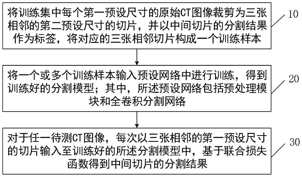
[0039] In order to solve the technical problems such as the strong subjectivity and empirical nature of traditional liver tumor image segmentation, the embodiment of the present invention provides a liver tumor segmentation method under CT images, such as figure 1 As shown, it mainly includes the following steps:

[0040] Step 10: Crop each original CT image of the first preset size in the training set into three adjacent slices of the second preset size, and use the segmentation result of the middle slice as a label to form a training sample from the corresponding three adjacent slices .

[0041] This step is mainly the construction of training samples. Wherein, the first preset size is larger than the second preset size; for example, the first preset size may be 512×512, and the second preset size may be 224×224. The specific implementation process is as follows: according to the liver and tumor annotations provided in the training set, the pixel coordinates of the liver a...

Embodiment 2

[0063] On the basis of the above-mentioned embodiment 1, the embodiment of the present invention further provides a liver tumor segmentation device under CT images, which can be used to realize the segmentation method in the embodiment 1. Such as Figure 5 As shown, the segmentation device mainly includes a sample generation module, a model training module and an image segmentation module.

[0064] The sample generation module is used to crop each original CT image of the first preset size in the training set into three adjacent slices of the second preset size, and take the segmentation result of the middle slice as a label, and divide the corresponding three adjacent slices constitute a training sample. For a more specific implementation process, reference may be made to step 10 in Embodiment 1, which will not be repeated here.

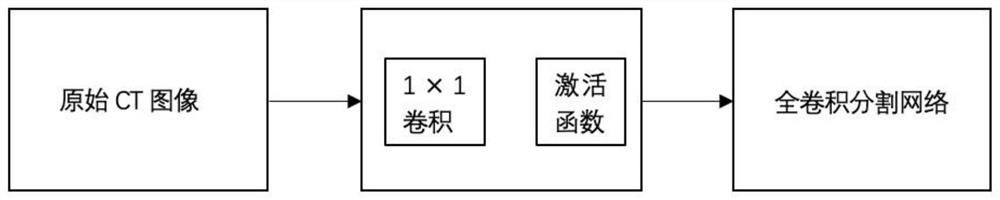
[0065] The model training module is used to input one or more training samples into a preset network for training to obtain a trained segmentatio...


PUM


Abstract

The invention discloses a method and a device for segmenting a liver tumor under a CT image. The method comprises the steps: cutting each original CT image of a first preset size in a training set into three adjacent slices of a second preset size, taking a segmentation result of a middle slice as a label, and enabling the corresponding three adjacent slices to form a training sample; inputting one or more training samples into a preset network for training to obtain a trained segmentation model; wherein the preset network comprises a preprocessing module and a full-convolution point cutting network; and for any to-be-detected CT image, inputting three adjacent slices of a first preset size into the trained segmentation model each time, and obtaining a segmentation result of a middle slice based on a joint loss function. According to the scheme, the problems existing in traditional liver tumor image segmentation can be effectively solved, the problems of large imaging difference of liver CT images, different tumor shapes and sizes and the like are solved, and the performance and effect of the automatic liver tumor segmentation method are improved.

Description

technical field [0001] The invention belongs to the technical field of image segmentation, and more specifically relates to a method and device for segmenting liver tumors under CT images. Background technique [0002] The liver is one of the essential organs of the human body and has functions such as metabolism and detoxification. Liver cancer is currently one of the most common cancers in the world, which seriously threatens human life and health. With the development of medical imaging technology, imaging examinations such as computed tomography (abbreviated as CT) and magnetic resonance imaging have been widely used in clinical diagnosis, among which CT technology is the most commonly used means in the examination and diagnosis of liver tumors . At present, the treatment methods for liver tumors mainly include surgical resection, ablation therapy, radiation therapy, etc., but no matter what kind of treatment method is used, it is necessary to accurately understand the ...

Claims


Application Information

Patent Timeline
IPC IPC(8): G06T7/10G06N3/08G06N3/04
CPCG06T7/10G06N3/08G06T2207/10081G06T2207/30056G06T2207/20081G06N3/045
Inventor 尤新革彭勤牧黄子轩朱文强
Owner HUAZHONG UNIV OF SCI & TECH RES INST SHENZHEN
Why Eureka
Try Eureka