Liver CT image tumor segmentation method based on information fusion

A CT image and CT image technology, applied in the field of image processing, can solve problems such as tumor edge blur, and achieve the effect of improving accuracy and reducing computational complexity

Pending Publication Date: 2021-11-16
XIAN UNIV OF TECH
View PDF0 Cites 0 Cited by
  • Summary
  • Abstract
  • Description
  • Claims
  • Application Information

AI Technical Summary

Problems solved by technology

[0005] The purpose of the present invention is to provide a liver CT image tumor segmentation method based on information fusion, which solves the problem of fuzzy tumor edge detection in the CT image tumor segmentation method existing in the prior art

Method used


Image

  • Liver CT image tumor segmentation method based on information fusion
  • Liver CT image tumor segmentation method based on information fusion
  • Liver CT image tumor segmentation method based on information fusion

Examples


Embodiment Construction

[0052] The present invention will be described in detail below with reference to the accompanying drawings and specific embodiments.

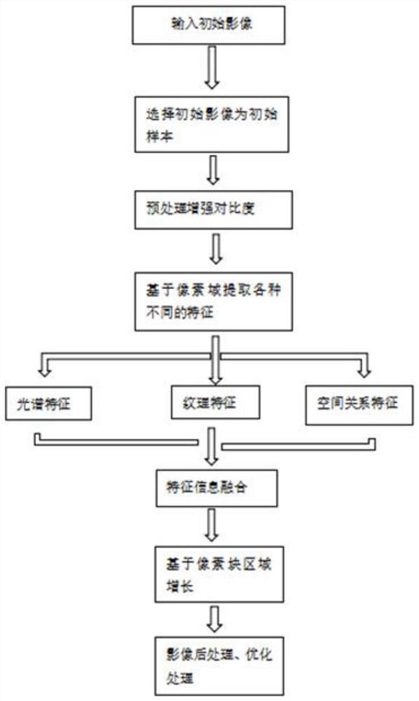
[0053] The present invention provides a liver CT image tumor segmentation method based on information fusion, such as figure 1 shown, follow the steps below:

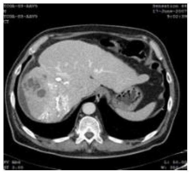
[0054] Step 1. Take the LiTS data set released by MICCAI as the experimental data set, take any liver CT image A from this data set, and the liver CT image A is used as input, and perform smoothing and denoising preprocessing on the image through the three-dimensional histogram reconstruction model. Obtain the preprocessed liver CT image B, such as figure 2 shown;

[0055] Step 1 is implemented according to the following steps:

[0056] Use the three-dimensional histogram reconstruction model to eliminate the gray level inhomogeneity between the pixel values ​​in the liver CT image A, and perform smoothing and denoising preprocessing on the liver CT image A to eliminate the noise in ...


PUM


Abstract

The invention discloses a liver CT image tumor segmentation method based on information fusion. The method specifically comprises the following steps: step 1, carrying out the smoothing and denoising preprocessing of an image through a three-dimensional histogram reconstruction model to obtain a preprocessed liver CT image B; step 2, respectively extracting three different features of spectrum, texture and spatial relationship of the liver CT image B to obtain three feature images corresponding to the liver CT image B; step 3, carrying out aggregation processing on the pixels of the three different feature images of the liver CT image B, and detecting the edge of the tumor part in the original image A by using an improved region growing algorithm to obtain a fused image C1; and step 4, performing post-processing operation on the edge of the tumor part detected in the image C1 fused in the step 3 in combination with morphological operation to form a final image segmentation result graph A. The method solves the problem of fuzzy tumor edge detection in a CT image tumor segmentation method in the prior art.

Description

technical field [0001] The invention belongs to the technical field of image processing, and in particular relates to a liver CT image tumor segmentation method based on information fusion. Background technique [0002] Liver image tumor segmentation technology is of great significance in disease judgment and preoperative prevention. The full name of CT is computed tomography. Compared with other imaging technologies, CT imaging has a higher density resolution, which improves the detection rate of lesions and the accuracy of diagnosis. CT imaging technology can not only diagnose brain diseases, but also examine the abdominal and thoracic organs of the human body to determine the condition of tumors, which can be used for the detection of various tissues and organs such as liver, lung, pancreas, and brain. However, due to the large variability of tissue features in medical images obtained by computed tomography and other imaging techniques, and the blurred boundary between s...

Claims


Application Information

Patent Timeline
Patent Type & Authority Applications(China)
IPC IPC(8): G06T7/11G06T7/13G06T7/44G06T5/00G06T5/30G06T5/40G06T5/50
CPCG06T7/11G06T7/13G06T5/40G06T5/50G06T7/44G06T5/30G06T2207/10081G06T2207/30056G06T5/70
Inventor 赵明华闫茹萍都双丽胡静石程李鹏石争浩
Owner XIAN UNIV OF TECH
Why Eureka
Try Eureka