Automatic Segmentation Method of 3D Liver CT Image Based on Supervoxel and Graph Cut Algorithm

A graph cut algorithm and CT image technology, applied in the field of medical image processing, can solve problems such as slow segmentation speed, high calculation amount, and complex calculation, so as to avoid the influence of algorithm robustness, reduce computational complexity, and have a high level of automation Effect

Inactive Publication Date: 2018-01-19
BEIJING UNIV OF TECH
View PDF4 Cites 0 Cited by
  • Summary
  • Abstract
  • Description
  • Claims
  • Application Information

AI Technical Summary

Problems solved by technology

The region growing algorithm has the advantages of being fast and easy to implement, but it is easy to cause inaccurate segmentation results when the gray level of the liver tissue is uneven
Segmentation algorithms based on active contours and level sets need to provide initial contours, and the calculation is complex and the segmentation speed is slow
Although the model-based segmentation algorithm can obtain more accurate segmentation results, the generation of probability maps or statistical shape models requires a large number of training images and corresponding manual segmentation standards, and the segmentation results may be inaccurate when dealing with non-standard shape livers
The graph cut algorithm is widely used in medical image segmentation because it can obtain the global optimal solution. However, directly constructing a graph in units of voxels for cutting will lead to excessive calculation and cannot obtain satisfactory segmentation results.

Method used


Image

  • Automatic Segmentation Method of 3D Liver CT Image Based on Supervoxel and Graph Cut Algorithm
  • Automatic Segmentation Method of 3D Liver CT Image Based on Supervoxel and Graph Cut Algorithm
  • Automatic Segmentation Method of 3D Liver CT Image Based on Supervoxel and Graph Cut Algorithm

Examples


Embodiment Construction

[0047] The extraction process is described in detail with reference to the accompanying drawings and practical examples. The image data used come from the enhanced CT scan images of the abdomen in the MICCAI2007Workshop database. The average size of each CT image is 512*512*208 pixels, and the average resolution is 0.68*0.68*1.6 mm.

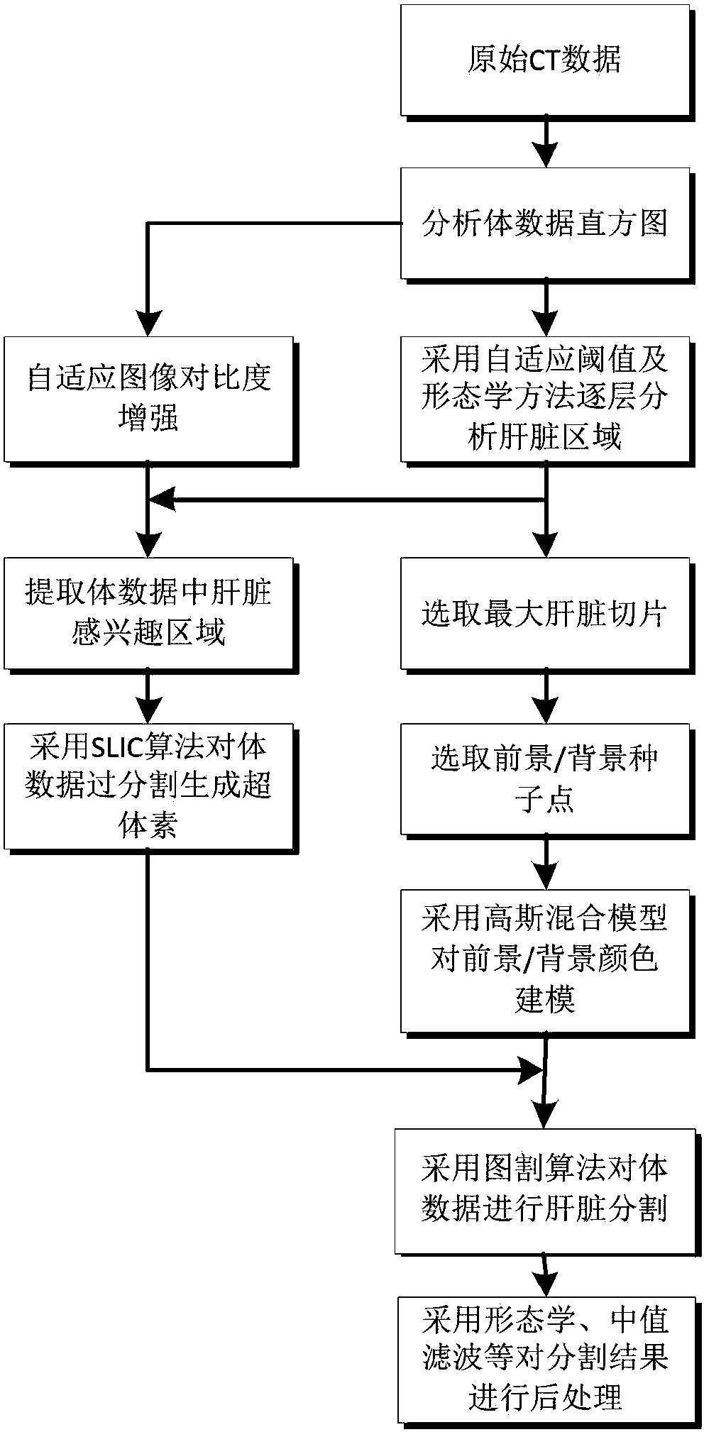
[0048] The flow chart of the liver CT image automatic segmentation method based on supervoxel and graph cut algorithm of the present invention is as follows figure 1 shown, including the following steps:

[0049] Step 1, for an input abdominal CT image I (such as Figure 4 shown) to perform histogram analysis, adaptively enhance the image contrast, and obtain the CT image I' after enhancing the contrast (such as Figure 5 shown). The specific implementation steps are as follows:

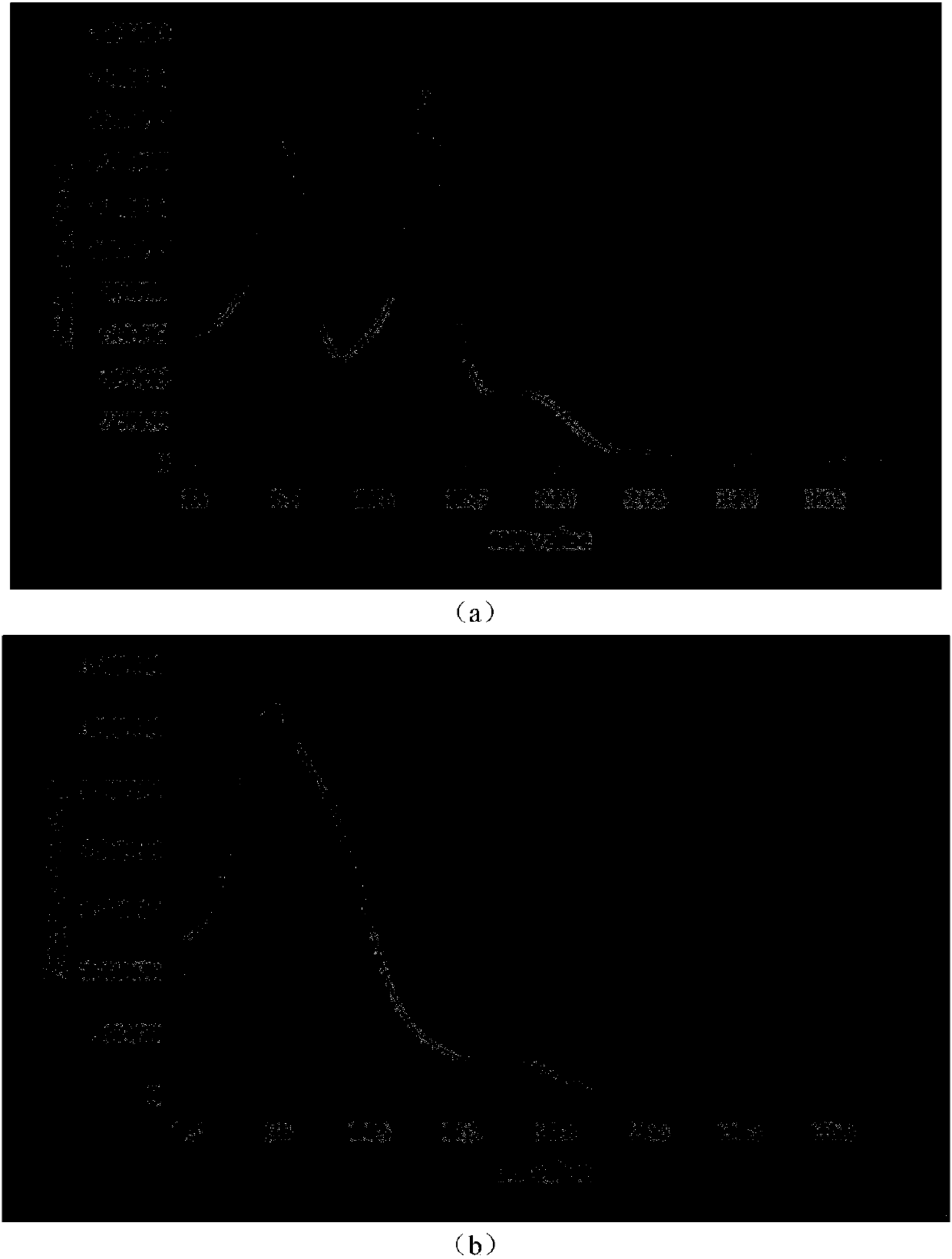
[0050] 1.1. Analyze the number of peaks in the image histogram. If there are two obvious peaks, it is a high-contrast image I high (Such as figure 2 As shown in...


PUM


Abstract

An automatic segmentation method for 3D liver CT images based on supervoxel and graph cut algorithm, through analyzing volume data histogram, and adaptively enhancing image contrast. Adaptive threshold and morphological methods were used to segment the initial contour of the liver layer by layer, and the largest liver slice was selected to calculate and extract the liver region of interest. In the largest liver slice, the seed points were selected according to the initial outline of the liver, and the Gaussian mixture model was used to model the foreground and background colors. The SLIC clustering algorithm was used to generate supervoxels for the contrast-enhanced liver region of interest, and an undirected weighted graph was constructed with the supervoxels as vertices, and the graph was cut using the graph cut algorithm. Finally, morphological operations are used to post-process the segmentation results. The invention can realize rapid and accurate automatic segmentation of the liver in the three-dimensional abdominal CT image.

Description

technical field [0001] The invention relates to the field of medical image processing, in particular to a three-dimensional liver CT image automatic segmentation method based on supervoxel and graph cut algorithms. Background technique [0002] Primary liver cancer is one of the most common malignant tumors in the world, with high morbidity and mortality. Computed Tomography (CT) imaging technology is widely used in the diagnosis and treatment of liver cancer due to its advantages of accurate anatomical information, high resolution, short scanning time and high penetration rate. Accurate 3D segmentation of the liver is a fundamental work in computer-aided diagnosis and an important prerequisite for 3D visualization, quantitative analysis, surgical planning, etc. At present, clinical liver segmentation is generally done manually by doctors based on experience, which is not only time-consuming and laborious, but also the accuracy varies from person to person. Therefore, effi...

Claims


Application Information

Patent Timeline
Patent Type & Authority Patents(China)
IPC IPC(8): G06T7/00G06T7/11G06T7/136G06T7/155
Inventor 吴薇薇周著黄吴水才白燕萍
Owner BEIJING UNIV OF TECH
Why Eureka
Try Eureka