
A Fusion Method of Hepatic Multiphase CT Images

A CT image and fusion method technology, applied in the field of medical image fusion, to achieve the effect of improving registration speed, improving accuracy, and improving mutual information measurement

Inactive Publication Date: 2018-06-08
XIAMEN UNIV
View PDF6 Cites 1 Cited by
  • Summary
  • Abstract
  • Description
  • Claims
  • Application Information

AI Technical Summary

Problems solved by technology

According to the characteristics of liver CT images, the present invention combines the image segmentation and image registration process into the image fusion process, thereby greatly improving the fusion accuracy

Method used


Image

  • A Fusion Method of Hepatic Multiphase CT Images
  • A Fusion Method of Hepatic Multiphase CT Images
  • A Fusion Method of Hepatic Multiphase CT Images

Examples


Embodiment Construction

[0076] The technical solution of the present invention will be further described in detail below in conjunction with the accompanying drawings and specific embodiments:

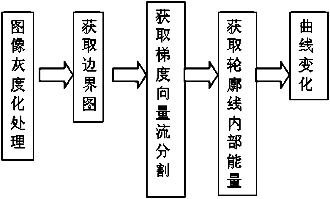
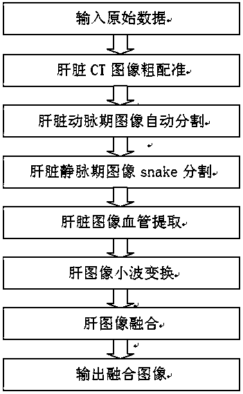
[0077] Such as figure 1 Shown: a liver multi-phase phase CT image fusion method, comprising the following steps:

[0078] Step 1, input arterial phase image sequence A and venous phase image sequence B;

[0079] 1a) storing the arterial phase image sequence A and the venous phase image sequence B to be fused into the hard disk of the computer respectively;

[0080] 1b) Respectively read the arterial phase image sequence A and the venous phase image sequence B to be fused stored in the hard disk space of the computer in step 1a).

[0081] Step 2, use the multi-resolution CT image registration method based on the joint histogram to perform rough registration, and for each CT image in the arterial phase image sequence A, find the CT image with the same spatial position in the venous phase image sequence B. Sp...


PUM


Abstract

The invention discloses a liver multi-phase CT image fusion method. First, a multi-resolution CT image registration method based on joint histograms is used to roughly register the source image sequence, and then combined with a region growing algorithm of confidence connection to realize liver The automatic image segmentation and the liver image segmentation based on the gradient vector flow snake model effectively extract the edge information of the liver; then the liver image is subjected to blood vessel extraction based on the directional region growing algorithm, and then the liver parenchyma image is subjected to free deformation based on B-spline Transformation and liver non-rigid registration based on spatial weighted mutual information to accurately find image pairs at the same location in space; finally, image fusion is performed based on wavelet transform. In view of the characteristics of liver CT images, the present invention combines the image segmentation and image registration processes into the image fusion process, thereby greatly improving the fusion accuracy.

Description

technical field [0001] The invention relates to the technical field of medical image fusion, in particular to a liver multiphase CT image fusion method. Background technique [0002] Image fusion refers to the image processing of image data about the same target collected from multiple sources or multiple times, extracting the respective beneficial information to the maximum extent, and finally synthesizing them into high-quality images, which is conducive to subsequent analysis. In the field of medical image processing, in order to obtain a more comprehensive, clearer, and more distinctive image of a certain part of the human body, image fusion is often used to improve image quality. This technology analyzes several groups of images together, synthesizes images with different details into an image with complete details, and keeps the details similar to the original image, so as to provide assistance for subsequent medical diagnosis. When performing image fusion, the images...

Claims


Application Information

Patent Timeline
Patent Type & Authority Patents(China)
IPC IPC(8): G06T3/00G06T3/40G06T7/00G06T7/11G06T7/136G06T5/50
CPCG06T3/4007G06T5/50G06T7/11G06T2207/30056G06T3/14
Inventor 黄晓阳王博亮周斌
Owner XIAMEN UNIV
Learn More