Human body part segmentation method and system based on SPECT imaging

A technology of human body parts and imaging, applied in the field of medical imaging, can solve problems such as difficulties, radiation pollution, and low signal-to-noise ratio between bones and backgrounds, and achieve the effects of precise segmentation, improved segmentation accuracy, and reduced calculation amount.

Active Publication Date: 2021-06-11
NORTHWEST UNIVERSITY FOR NATIONALITIES
View PDF8 Cites 6 Cited by
  • Summary
  • Abstract
  • Description
  • Claims
  • Application Information

AI Technical Summary

Problems solved by technology

The principle of SPECT imaging limits the processing and analysis of traditional methods based on classification and segmentation in whole-body bone scan imaging. In SPECT imaging, the difference between the radiation value of human bones and the background value is small, which makes it very easy for bone and background to occur during imaging. The adhesion phenomenon, and the existence of external radiation pollution, which makes the image of human bones unclear in SPECT imaging, the signal-to-noise ratio of bones and background is low, the boundary between bones and soft tissues is not obvious, SPECT between different patients of different ages Imaging variability and patient morphology on SPECT scans make SPECT imaging analysis difficult based on computer-aided diagnostic techniques
At present, no method has been found that can effectively segment the head, trunk, limbs, pelvis, etc. for each SPCET whole-body bone imaging data matrix

Method used


Image

  • Human body part segmentation method and system based on SPECT imaging
  • Human body part segmentation method and system based on SPECT imaging
  • Human body part segmentation method and system based on SPECT imaging

Examples


Embodiment 1

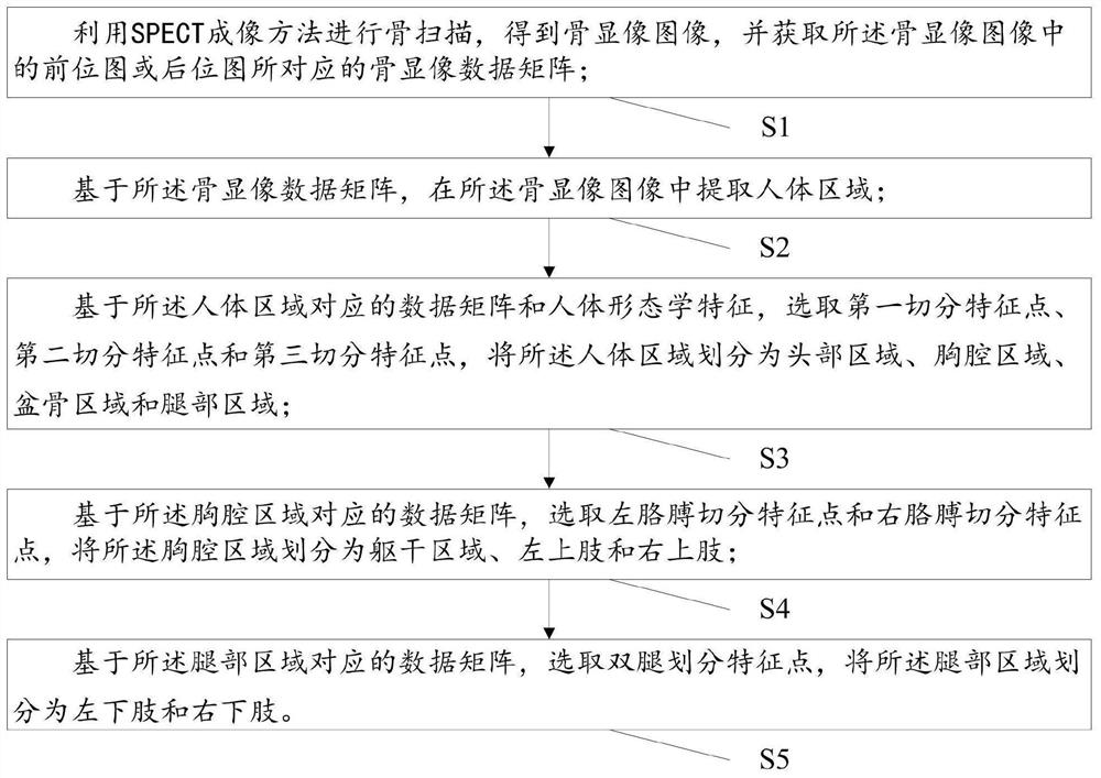
[0042] The existing technology is mainly aimed at human body segmentation of traditional natural images, which not only has high algorithm complexity, but also cannot be directly applied to bone imaging images. In the segmentation of whole-body bone imaging images, an accurate human body segmentation method that can effectively target each whole-body bone imaging image has not yet been found. This embodiment is used to provide a human body part segmentation method based on SPECT imaging, which can be applied to the human body segmentation of each bone imaging image, such as figure 1 As shown, the segmentation method includes the following steps:

[0043] S1: performing a bone scan using the SPECT imaging method to obtain a bone imaging image, and acquiring a bone imaging data matrix corresponding to the anterior or posterior image in the bone imaging image;

[0044] Specifically, S1 includes: capturing the nuclide amount by a nuclide detector, displaying it as a bone imaging ...

Embodiment 2

[0097] This embodiment is used to provide a human body part segmentation system based on SPECT imaging, such as Figure 15 As shown, the segmentation system includes:

[0098] The bone imaging data matrix acquisition module M1 is used to perform bone scanning using the SPECT imaging method to obtain a bone imaging image, and acquire the bone imaging data matrix corresponding to the anterior bitmap or the posterior bitmap in the bone imaging image ;

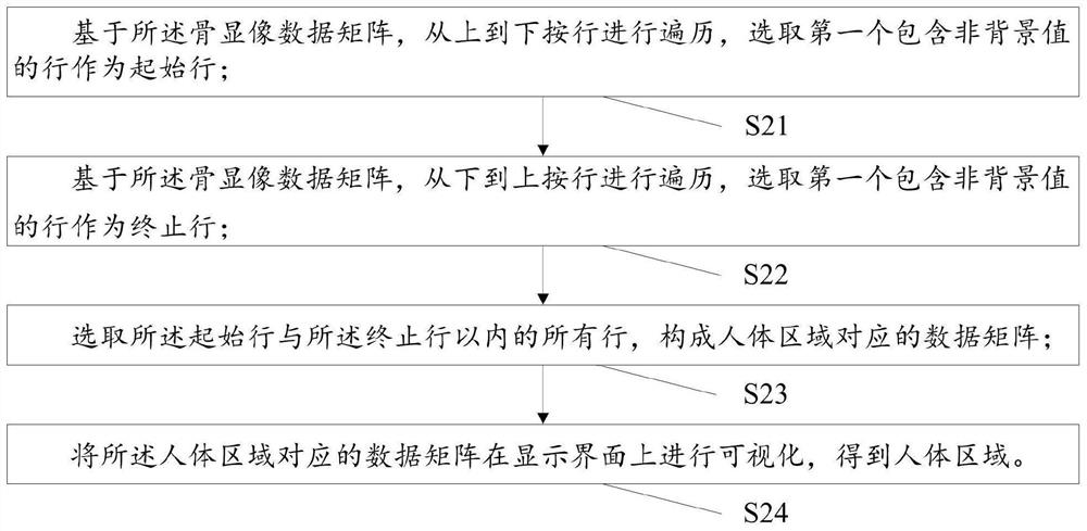
[0099] A human body area extraction module M2, configured to extract a human body area in the bone imaging image based on the bone imaging data matrix;

[0100] The human body area division module M3 is used to select the first segmentation feature point, the second segmentation feature point and the third segmentation feature point based on the data matrix corresponding to the human body area and the human body morphological features, and divide the human body area Divided into head region, thoracic region, pelvic region and le...


PUM


Abstract

The invention relates to a human body part segmentation method and system based on SPECT imaging, based on a bone imaging data matrix, a human body region is extracted from a bone imaging image to obtain effective information of the bone imaging data matrix, the calculation amount is reduced, and the segmentation precision is improved. Then the human body area is divided into a head area, a chest area, a pelvic bone area and a leg area based on the data matrix corresponding to the human body area and human morphological characteristics, then the chest area is divided into a trunk area, a left upper limb and a right upper limb based on the data matrix corresponding to the chest area, and finally based on the data matrix corresponding to the leg area, the leg area is divided into the left lower limb and the right lower limb, so that the human body is divided into the head, the trunk, the four limbs, the pelvis and other parts, accurate segmentation of the human body parts is achieved, and accurate segmentation of the head, the trunk, the four limbs, the pelvis and other parts can be effectively conducted on any SPCET bone imaging image.

Description

technical field [0001] The invention relates to the technical field of medical imaging, in particular to a method and system for segmenting human body parts based on SPECT imaging. Background technique [0002] Medical imaging is an important technical means for the diagnosis and treatment of modern clinical diseases. It can capture and present the internal structure and functional status of the body in a non-invasive way. As a general term, medical imaging usually includes two relatively independent components: medical imaging and medical image processing. [0003] In the field of medical imaging, nuclear medicine imaging is regarded as an effective medical method for diagnosis, treatment, evaluation and prevention of various serious diseases. Since the British physicist Roentgen discovered X-rays in 1895, medical imaging has developed from traditional structural imaging modalities such as computerized tomography (CT), magnetic resonance imaging (Magnetic Resonance Imaging...

Claims


Application Information

Patent Timeline
Patent Type & Authority Applications(China)
IPC IPC(8): G06T7/00G06K9/00G06K9/34A61B6/03
CPCG06T7/0012A61B6/037G06T2207/10108G06T2207/30008G06V40/10G06V10/267
Inventor 林强李同同满正行曹永春
Owner NORTHWEST UNIVERSITY FOR NATIONALITIES
Why Eureka
Try Eureka