Quantitative measurement and analysis system for temporal-mandibular joint on magnetic resonance image

A temporomandibular joint and magnetic resonance image technology, applied in the field of quantitative measurement and analysis of the upper temporomandibular joint, can solve the problems of condylar head shape variability, offset, measurement error, etc., and achieve the effect of wide range consistency

Pending Publication Date: 2021-06-22
SHANGHAI NINTH PEOPLES HOSPITAL AFFILIATED TO SHANGHAI JIAO TONG UNIV SCHOOL OF MEDICINE
View PDF0 Cites 0 Cited by
  • Summary
  • Abstract
  • Description
  • Claims
  • Application Information

AI Technical Summary

Problems solved by technology

[0005] like figure 1 As shown, the measurement system referenced by the condyle’s mental position is convenient for evaluating the height, width, positional relationship between the articular disc and ...

Method used


Image

  • Quantitative measurement and analysis system for temporal-mandibular joint on magnetic resonance image
  • Quantitative measurement and analysis system for temporal-mandibular joint on magnetic resonance image
  • Quantitative measurement and analysis system for temporal-mandibular joint on magnetic resonance image

Examples


Embodiment 1

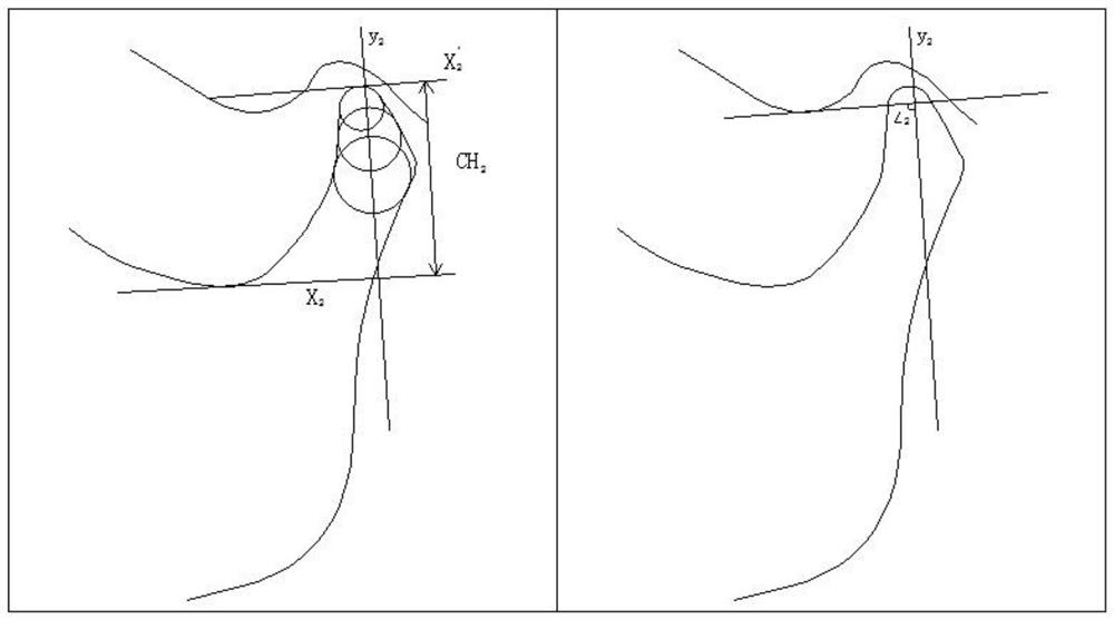
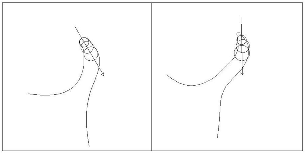
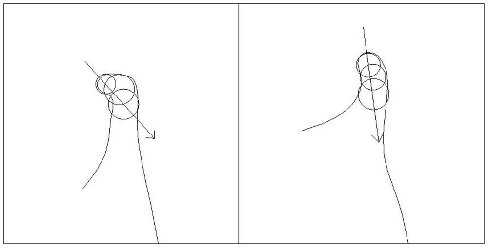
[0034] see Figure 4-9 According to an embodiment of the present invention, a magnetic resonance image upper temporomandibular joint quantitative measurement and analysis system includes the following steps:

[0035] S101, such as Figure 4 As shown, in the MRI image, the largest cross-sectional image of the condyle is used as the measurement object, and the shape of the condyle and mandibular ramus is depicted, and the most protruding point of the condyle and the most protruding point of the mandibular angle are selected at the posterior edge of the mandibular ramus. Connect the two points to form a tangent line to the posterior edge of the mandibular ramus, which is the vertical reference line.

[0036] S103, such as Figure 5 As shown, vertically tangent to the posterior edge of the mandibular ramus, make a straight line tangent to the sigmoid notch of the mandible, and determine the horizontal reference line.

[0037] S105, in the direction of the vertical reference lin...


PUM


Abstract

The invention discloses a quantitative measurement and analysis system for temporal-mandibular joints on a magnetic resonance image. The system comprises the following steps: step 1, taking a condyle process maximum cross-section image in an MRI image as a measurement object, describing condyle process and mandibular ramus forms, selecting a most prominent point of the condyle process backwards and a most prominent point of the mandibular ramus backwards at the rear edge of the mandibular ramus, and connecting the two points to form a mandibular ramus trailing edge tangent line, namely a vertical reference line; and step 2, making a straight line perpendicular to the tangent line of the rear edge of the mandibular ramus to be tangent to the B-shaped incisura of the mandible, and determining a horizontal reference line. The system has the advantages that: a stable TMJ MRI measurement system can be established, the condylar process height can be effectively evaluated, comparison before and after treatment is carried out, and the treatment effect is evaluated; and the severity of the TMJ organic lesion can be evaluated by comprehensively evaluating the discoid-condylar distance, the joint discoid length and the condylar process height, so that formulation of a treatment scheme is guided, and the treatment effect is predicted.

Description

technical field [0001] The invention relates to the technical field of quantitative measurement and analysis of upper temporomandibular joints, in particular to a system for quantitative measurement and analysis of upper temporomandibular joints in magnetic resonance images. Background technique [0002] Magnetic resonance imaging (MRI) of the temporomandibular joint (TMJ) can effectively image the calcified and non-calcified tissues inside the joint at the same time, and is the most effective means to determine whether there is an abnormality in the internal structure of the joint; Radiation damage, so it is considered safer than T. TMJ MRI is also an indispensable measure in disease diagnosis, prognosis judgment and curative effect evaluation; and many patients do not need to undergo T examination during the diagnosis process. [0003] Therefore, it is very important to obtain quantitative TMJ evaluation indicators from MRI. During measurement, the setting of the referen...

Claims


Application Information

Patent Timeline
IPC IPC(8): A61B5/055
CPCA61B5/055A61B5/0033
Inventor 谢千阳杨驰陈敏洁白果刘芷扬李佩伦
Owner SHANGHAI NINTH PEOPLES HOSPITAL AFFILIATED TO SHANGHAI JIAO TONG UNIV SCHOOL OF MEDICINE
Why Eureka
Try Eureka