Retinal vessel segmentation method and device based on multi-scale attention network, and storage medium

A retinal blood vessel, attention technology, applied in neural learning methods, biological neural network models, image analysis, etc., can solve the problem of ignoring specific areas of scale changes

Inactive Publication Date: 2021-06-22
SHANDONG UNIV
View PDF2 Cites 3 Cited by
  • Summary
  • Abstract
  • Description
  • Claims
  • Application Information

AI Technical Summary

Problems solved by technology

The great success of the deep convolutional neural network CNN has made U-Net widely used in medical image segmentation, but the network also ignores the proportional changes in the path and specific regions in the feature map, and the segmentation accuracy has a further improvement. room for improvement

Method used


Image

  • Retinal vessel segmentation method and device based on multi-scale attention network, and storage medium
  • Retinal vessel segmentation method and device based on multi-scale attention network, and storage medium
  • Retinal vessel segmentation method and device based on multi-scale attention network, and storage medium

Examples


Embodiment 1

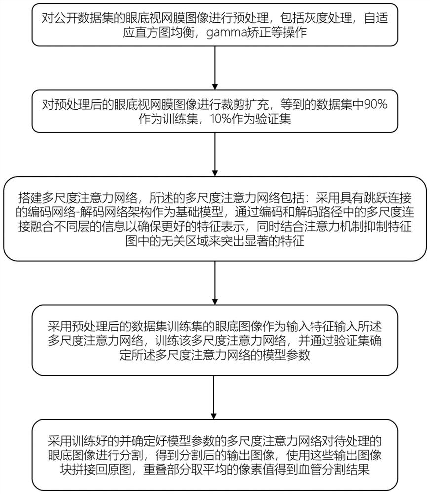
[0070] A retinal vessel segmentation method based on Multi-Scale AttentionNet, such as figure 1 shown, including the following steps:

[0071] Step 1: Get the dataset;

[0072] Step 2: Dataset preprocessing;

[0073] Perform grayscale processing, adaptive histogram equalization, and gamma correction on the data set obtained in step 1;

[0074] Step 3: Construct a retinal vessel segmentation model;

[0075] Step 4: Training the retinal vessel segmentation model; refers to: use the PyTorch framework to train the retinal vessel segmentation model, the loss function uses the combination of the cross entropy loss function and the dice loss function to solve the class balance problem, the coefficient is 0.5, and the learning rate is set is 0.0002 and the number of iterations is 150.

[0076] Step 5: retinal vessel segmentation test;

[0077] Preprocessing the fundus retinal image test set (sequentially performing grayscale processing, adaptive histogram equalization, and gamma ...

Embodiment 2

[0080] According to a kind of retinal blood vessel segmentation method based on multi-scale attention network (Multi-ScaleAttentionNet) described in embodiment 1, its difference is:

[0081] In step 2, data set preprocessing includes the following steps:

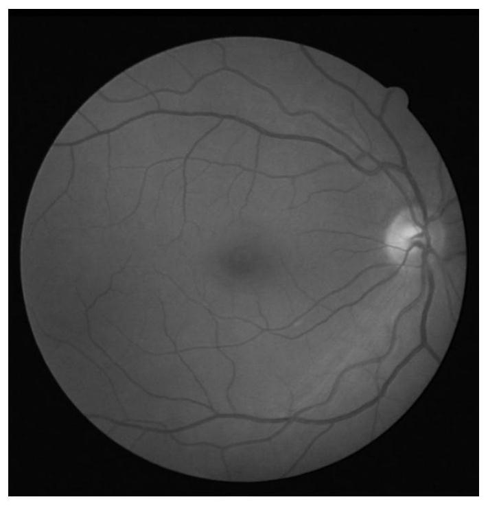
[0082] Step 2.1: Perform grayscale processing on the data set obtained in step 1, and convert all pictures into grayscale images; figure 2 It is a schematic diagram of the retinal grayscale image of tra_40, one of the pictures in the training set in the existing DRIVE dataset.

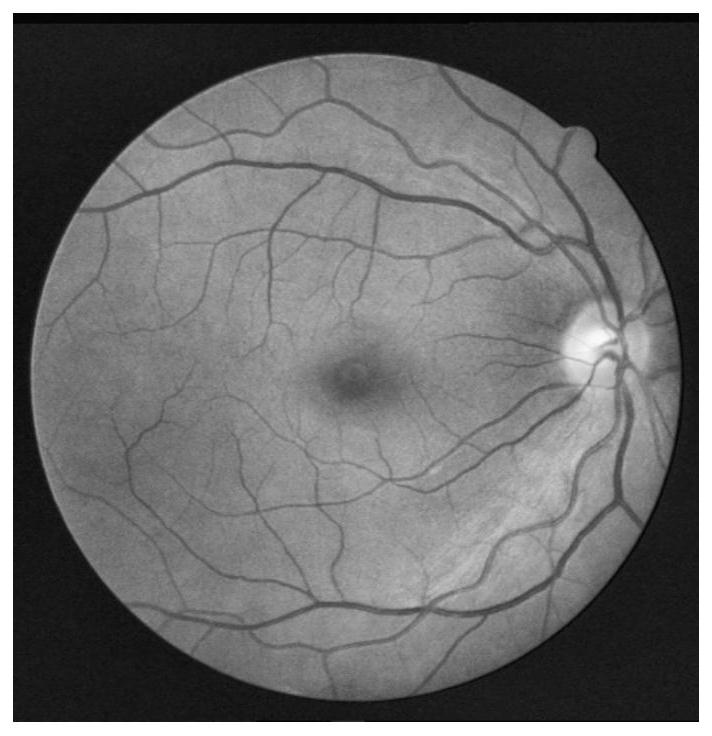
[0083] Step 2.2: Perform contrast-limited adaptive histogram equalization on the grayscale image obtained in step 2.1; the contrast-limited adaptive histogram equalization algorithm can effectively limit noise amplification.

[0084] Step 2.3: Use gamma correction to perform non-linear operations on the image obtained after processing in step 2.2. This will reduce the noise of the image and improve the overall contrast of blood vessels, which is b...

Embodiment 3

[0090] According to a kind of retinal blood vessel segmentation method based on multi-scale attention network (Multi-ScaleAttentionNet) described in embodiment 2, its difference is:

[0091] Such as Figure 5 As shown, the retinal vessel segmentation model includes a multi-scale connection network and an attention module;

[0092] The multi-scale connection network includes an encoding path and a decoding path. Both the encoding path and the decoding path include four spatially scaled blocks, and each spatially scaled block is passed through twice a 2D convolutional filter of size 3×3, ReLU, and batch normalization. (BN) layer, and the input and the output feature map of the input through the convolutional layer are connected; this will reduce overfitting and reduce the size of the input by half, which is beneficial for the network to learn contextual information.

[0093] In the encoding path, in each spatial scale block, the input and the output feature map of the input thr...


PUM


Abstract

The invention relates to a retinal vessel segmentation method and device based on a multi-scale attention network, and a storage medium. The method comprises the steps: 1, obtaining a data set; 2, preprocessing the data set, and sequentially carrying out gray processing, adaptive histogram equalization and gamma correction processing on the data set; 3, constructing a retinal vessel segmentation model; 4, training a retinal vessel segmentation model; 5, performing retinal blood vessel segmentation: preprocessing a to-be-segmented fundus retina image and then inputting the to-be-segmented fundus retina image into the trained retinal vessel segmentation model to obtain a segmented output image; 6, splicing the segmented output images to obtain an original image, and taking an average pixel value of overlapped parts to obtain a retinal blood vessel segmentation result. According to the method, the feature maps of all the layers are fused, and better feature representation is obtained. An attention module is added, which pays attention to those areas that contribute more to the result to obtain a more accurate result.

Description

technical field [0001] The invention relates to deep learning algorithms in the field of medical image processing, in particular to a retinal vessel segmentation method, device and storage medium based on a multi-scale attention network. Background technique [0002] As one of the most important organs of the human body, the eye plays a vital role in human observation and cognition of the world. Protecting human vision has always been a hot spot of widespread concern in society. As China is the country with the largest number of blind people in the world, it is necessary to take practical and effective measures to prevent blindness. Fundus disease is the main factor leading to irreversible blindness in my country, so it is necessary to carry out large-scale fundus screening and conduct regular fundus retinal examinations for patients with potential fundus diseases, which is very important for active prevention, early diagnosis and treatment of fundus diseases significance. ...

Claims


Application Information

Patent Timeline
Patent Type & Authority Applications(China)
IPC IPC(8): G06T7/11G06T5/40G06T5/00G06N3/08
CPCG06T7/11G06T5/40G06T5/002G06T5/006G06T5/007G06N3/08G06T2207/20081G06T2207/20084G06T2207/20132G06T2207/30041G06T2207/30101
Inventor 刘安琪吴雨林周洪超
Owner SHANDONG UNIV
Why Eureka
Try Eureka