U-Net kidney tumor image segmentation method and device based on aggregation interlayer information, and storage medium

A kidney tumor and image segmentation technology, applied in image analysis, image enhancement, image coding, etc., can solve the problems of complex 3D model structure, prone to overfitting, and high hardware requirements, so as to improve the segmentation accuracy and reduce hardware. Requires, enhances the effect of features

Inactive Publication Date: 2021-06-22
SHANDONG UNIV
View PDF6 Cites 1 Cited by
  • Summary
  • Abstract
  • Description
  • Claims
  • Application Information

AI Technical Summary

Problems solved by technology

However, the structure of the 3D model is relatively complex, there are many parameters, it is prone to overfitting, and the demand for hardware is high

Method used


Image

  • U-Net kidney tumor image segmentation method and device based on aggregation interlayer information, and storage medium
  • U-Net kidney tumor image segmentation method and device based on aggregation interlayer information, and storage medium
  • U-Net kidney tumor image segmentation method and device based on aggregation interlayer information, and storage medium

Examples


Embodiment 1

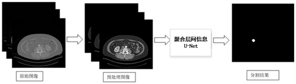
[0071] A U-Net kidney tumor image segmentation method based on aggregated inter-layer information, such as figure 2 shown, including the following steps:

[0072] 1) Image acquisition;

[0073] 2) Image preprocessing;

[0074] Set the window level and window width, remove the noise interference in the incoherent area of ​​the CT image obtained in step 1), and keep the brightness and contrast of the kidney tumor image consistent through histogram equalization, normalization and standardization in turn;

[0075] 3) Build tags;

[0076] Construct corresponding labels for the preprocessed images obtained in step 2); prepare for the training and testing of the input network in the next step. The number of input labels is 3 consecutive slices, only the slice in the center is the target slice, and the other two input slices are used as auxiliary slices to provide spatial information.

[0077] 4) Build a U-Net model that aggregates inter-layer information;

[0078] The U-Net mod...

Embodiment 2

[0088]A U-Net kidney tumor image segmentation method based on aggregated inter-layer information according to Embodiment 1, the difference is:

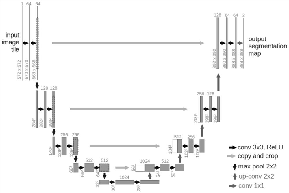
[0089] The U-Net model for aggregating inter-layer information is based on 3D networks and 2D networks. like Figure 4 As shown, the U-Net model that aggregates inter-layer information includes an encoder-decoder network, and the encoder-decoder network includes an encoder subnet and a decoder subnet. The encoder subnet performs feature extraction and applies 3D convolution to utilize Inter-layer information, the network uses the information provided by the encoder subnet to restore the image size and perform semantic segmentation in the decoder subnet; through the decoder subnet, 2D convolution is used to reduce the size of the network to predict only a certain slice;

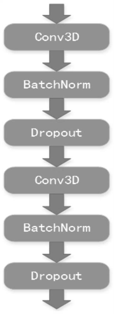
[0090] The encoder subnet is divided into four layers with a downsampling at the end of each layer; the resolution is decreased and the number of feature images is d...

Embodiment 3

[0101] The kidney tumor image segmentation method based on the U-net network of aggregated inter-layer information according to Embodiment 1 or 2, the difference is:

[0102] In step 1), image acquisition refers to: using ITK-SNAP to read the data set of the KiTS19 Challenge to acquire CT images. Figure 5 Image diagram for the KiTS19 Challenge dataset.

[0103] In step 1), image preprocessing aims to improve the precision and accuracy of the processing algorithm in the next stage, including:

[0104] A. Window truncation:

[0105] The window level is the center of the CT value range. If the CT value range is set to [a,b], the window level is equal to (a+b) / 2;

[0106] The window width represents a relative range, set as [a, b], which represents the CT value range of the image; when a tissue area is observed clinically, the window level will be set as the CT reference value of the tissue, and then different windows will be adjusted to adjust Wide, display, observe the condi...


PUM


Abstract

The invention relates to a U-Net kidney tumor image segmentation method and device based on aggregation interlayer information, and a storage medium. The method comprises the following steps: 1) acquiring an image; 2) image preprocessing; 3) constructing a label, and constructing a corresponding label for the preprocessed image obtained in the step 2); 4) constructing a U-Net model for aggregating interlayer information; 5) training a U-Net model for aggregating interlayer information; 6) segmenting the kidney tumor image, and inputting a kidney tumor image to be segmented into the trained U-Net model of the aggregation interlayer information to realize image segmentation. According to the method, the information in the slice layers and the information between the slice layers are fully utilized to learn context information, and memory consumption and hardware requirements of a computer are reduced. The output extraction features after convolution are realized on multiple scales, so the information redundancy can be reduced, and the segmentation accuracy can be improved.

Description

technical field [0001] The invention relates to a U-Net kidney tumor image segmentation method, equipment and storage medium based on aggregated inter-layer information, and belongs to the field of computer-aided technology. Background technique [0002] There are more than 400,000 new cases of kidney tumors each year, for which surgery is the most common treatment. Due to the morphological diversity of kidney and renal tumors, there is currently great interest in the impact of tumor morphology on surgery and the development of advanced surgical planning techniques, and image segmentation is an effective tool for doing so. [0003] In recent years, with the improvement of computer computing performance, it has become possible to achieve computer-aided segmentation of medical images. However, due to some difficulties in the segmentation of kidneys in CT images, for example, the contrast of CT images is low, the boundaries of kidney organs are relatively blurred, and the diffe...

Claims


Application Information

Patent Timeline
Patent Type & Authority Applications(China)
IPC IPC(8): G06T7/11G06T9/00G06K9/62G06N3/04G06N3/08
CPCG06T7/11G06T9/002G06N3/08G06T2207/10081G06T2207/20081G06T2207/20084G06T2207/20116G06T2207/30084G06T2207/30096G06N3/044G06N3/045G06F18/253
Inventor 王鹏伟初莹莹花蒨蒨张翅
Owner SHANDONG UNIV
Why Eureka
Try Eureka