[0032] In order to facilitate understanding of the present invention, the present invention will be described more fully below. However, the present invention may be embodied in many different forms and is not limited to the embodiments described herein. Rather, these embodiments are provided so that a thorough and complete understanding of the present disclosure is provided.
[0033] Unless otherwise defined, all technical and scientific terms used herein have the same meaning as commonly understood by one of ordinary skill in the art to which this invention belongs. The terms used herein in the description of the present invention are for the purpose of describing specific embodiments only, and are not intended to limit the present invention.
[0034] The technical solutions of the patent of the present invention will be clearly and completely described below. Obviously, the described embodiments are part of the embodiments of the present invention, but not all of the embodiments. Based on the embodiments of the present invention, all other embodiments obtained by those skilled in the art without creative efforts shall fall within the protection scope of the present invention.
[0035] The present invention provides an immune cell that autocrines IL-15 and anti-PD1 fusion protein, and uses gene-edited immune cells to secrete IL-15 and anti-PD1 fusion protein, so as to improve the activity of immune cells and improve the anti-tumor effect. Killing effect, immune cells that autocrine IL-15 and anti-PD1 fusion proteins include: fusion proteins expressed in series according to the sequence of anti-PD1, G4S*4 Linker, IL-15N72D, G4S*4 Linker and IL-15RaSu, and the expression The fusion protein and the gene-edited cells affected by the autocrine fusion protein.
[0036]Immune cells themselves do not express the fusion proteins mentioned above. In order for immune cells to secrete the fusion proteins, they need to undergo gene editing. In addition, cells used for tumor treatment such as CarT/CarNK/TCR-T/IPS are already genetically engineered. Edited and then made to secrete fusion proteins are still edited immune cells, so they are collectively referred to as gene-edited immune cells here. The autocrine IL-15 and anti-PD1 fusion protein immune cells of the present invention combine IL-15 and IL15Ra hyperkinesin and anti-PD1 fusion protein, and obtain chimeric antigen receptor T cells that successfully secrete the fusion protein (Chimeric antigen receptor CART).
[0037] anti-PD1 is anti-PD1 VL and/or anti-PD1 VH.
[0038] In one embodiment, the immune cells express a chimeric antigen receptor (CAR). The expression of the chimeric antigen receptor can be a CAR targeting one target, or a CAR targeting two targets or multiple targets.
[0039] In one embodiment, the target of the chimeric antigen receptor may be the idiotype of the anti-CLDN18.2 antibody.
[0040] The target of the chimeric antigen receptor can also be one or more of the idiotypes CLDN18.2, GPC3, HER2, TAA, GD2, MSLN, EGFR, NY-ESO-1, MUCl, PSMA and EBV.
[0041] The binding region of the chimeric antigen receptor and target can be scFv and/or Fab. In one embodiment, the structure of the scFv region in the chimeric antigen receptor structure can be replaced by any single-chain antibody, single-chain variable fragment (scFv) and Fab fragment of any target.
[0042] Chimeric antigen receptors contain a leader sequence, an scFv that recognizes tumor-associated antigens, a hinge and transmembrane domain, an intracellular co-stimulatory domain, and the intracellular activation signal CD3ζ.
[0043] scFv is the scFv of anti-idiotype antibody; hinge region and transmembrane domain are CD28 or CD8 hinge region and transmembrane domain; intracellular costimulatory domain is CD28 or CD137 (4-1BB) or ICOS intracellular costimulatory structure area.
[0044] The binding region of the chimeric antigen receptor and the target can be a bispecific antibody that binds to one target, or a bispecific antibody that binds two targets, or two or more chimeric antigen receptors are formed across the membrane respectively. and identify different targets.
[0045] The division between immune cells and autocrine IL-15 and anti-PD1 fusion protein is a protein cleavage functional element. The protein cleavage functional element can be T2A, P2A, E2A, F2A or IRES.
[0046] In one embodiment, the gene transfer method of immune cells is: lentivirus, retrovirus, common plasmid vector, episomal vector, nano-delivery system, electrotransduction, transposon or other delivery systems.
[0047] In one embodiment, the immune cells are T cells, NK cells, NKT cells, macrophages, gamma-delta T cells, TIL cells, TCR-T cells or other tumor killer cells.
[0048] Immune cells that autocrine IL-15 and anti-PD1 fusion protein can be made into preparations, and the preparations are pharmaceutically acceptable carriers, diluents or excipients. Administration of the formulations of the present invention can be carried out in any convenient manner, including by nebulization, injection, swallowing, infusion, implantation or transplantation. The formulation can be used for the prevention and/or treatment of solid tumors.
[0049] The autocrine IL-15 and anti-PD1 fusion protein immune cells of the present invention express CAR, which specifically recognizes the idiotype of the anti-autoantibody; the hyperkinesin and anti-PD1 fusion protein combined with IL-15 and IL15Ra, and The fusion protein is successfully secreted by the CART cells to enhance the proliferation ability, anti-apoptosis ability and tumor killing ability of the CarT cells; in addition, the immune cells of the present invention can specifically kill and secrete the fusion protein of IL-15 and anti-PD1; and The killing effect is precise, the safety is higher, it is not easy to recur, and the quality of life of patients is improved.
[0050] The following is a description of specific embodiments.
[0051] In the following examples, the preparation of CarT and the secretion of IL-15 and anti-PD1 fusion protein from T cells in peripheral blood is used as an example. The preparation method and functional verification of immune cells include the following steps:
[0052] 1) Structural design of fusion protein;
[0053] 2) Construct secreted CART cells and conduct in vitro functional tests;
[0054] 3) In vivo functional test of CART cells secreting fusion protein.
[0055] 1) Structural design of fusion protein
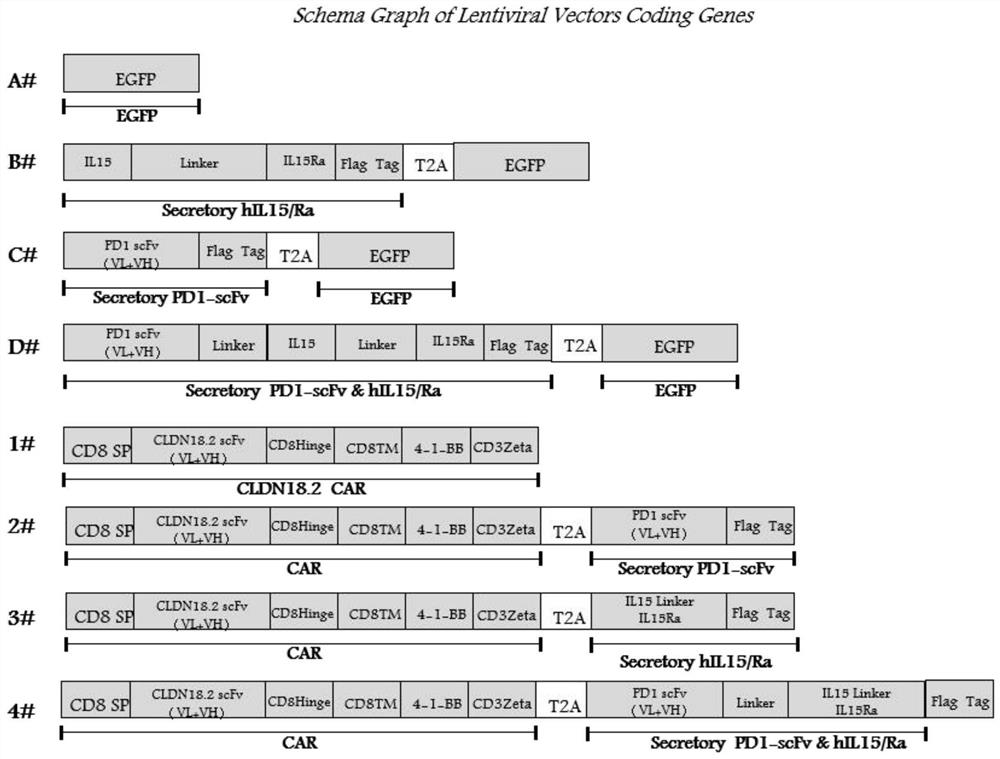
[0056] According to the sequence of hIL15, hIL15Ra, anti-PD1, according to figure 1 The middle structure was designed as the fusion protein structure in A#~D#, and it was designed into CarT-CLDN18.2, where 1# was the control CarT, and 2#~4# were secreted CarT.
[0057] Wherein, the amino acid sequence of hIL15 is: METDTLLLWVLLLWVPGSTGNWVNVISDLKKIEDLIQSMHIDATLYTESDVHPSCKVTAMKCFLLELQVISLESGDASIHDTVENLIILANDSLSSNGNVTESGCKECEELEEKNIKEFLQSFVHIVQMFINTS.
[0058] The amino acid sequence of hIL15Ra is: ITCPPPMSVEHADIWVKSYSLYSRERYICNSGFKRKAGTSSLTECVLNKATNVAHWTTPSLKCIR.
[0059] Anti-PD1 VH amino acid sequence is: QVQLVESGGGVVQPGRSLRLDCKASGITFSNSGMHWVRQAPGKGLEWVAVIWYDGSKRYYADSVKGRFTISRDNSKNTLFLQMNSLRAEDTAVYYCATNDDYWGQGTLVTVSS.
[0060] Anti-PD1 VL amino acid sequence is: EIVLTQSPATLSLSPGERATLSCRASQSVSSYLAWYQQKPGQAPRLLIYDASNRATGIPARFSGSGSGTDFTLTISSLEPEDFAVYYCQQSSNWPRTFGQGTKVEIK.
[0061] The amino acid sequence of Linker is: GSSSSGSSSSGSSSSGSSSS.
[0062] 2) Construction of secreted CART cells and in vitro functional tests
[0063] Construction of secreted CART cells and in vitro functional tests include the following steps:
[0064] S100: Cell Line Culture
[0065] The base sequence expressing CLDN18.2 was cloned into the backbone of PHBLV lentiviral vector and placed under the promoter of EF1α (EF-1α) to form PHBLV-EF1α-CLDN18.2, PHBLV-EF1α-CLDN18.2, slow The viral envelope plasmid pMD2.G (Addgene, Plasmid#12259) and the lentiviral packaging plasmid psPAX2 (Addgene Plasmid#12260) were transferred into 293T using Lipofectamine3000 to prepare a complete lentiviral expression vector;
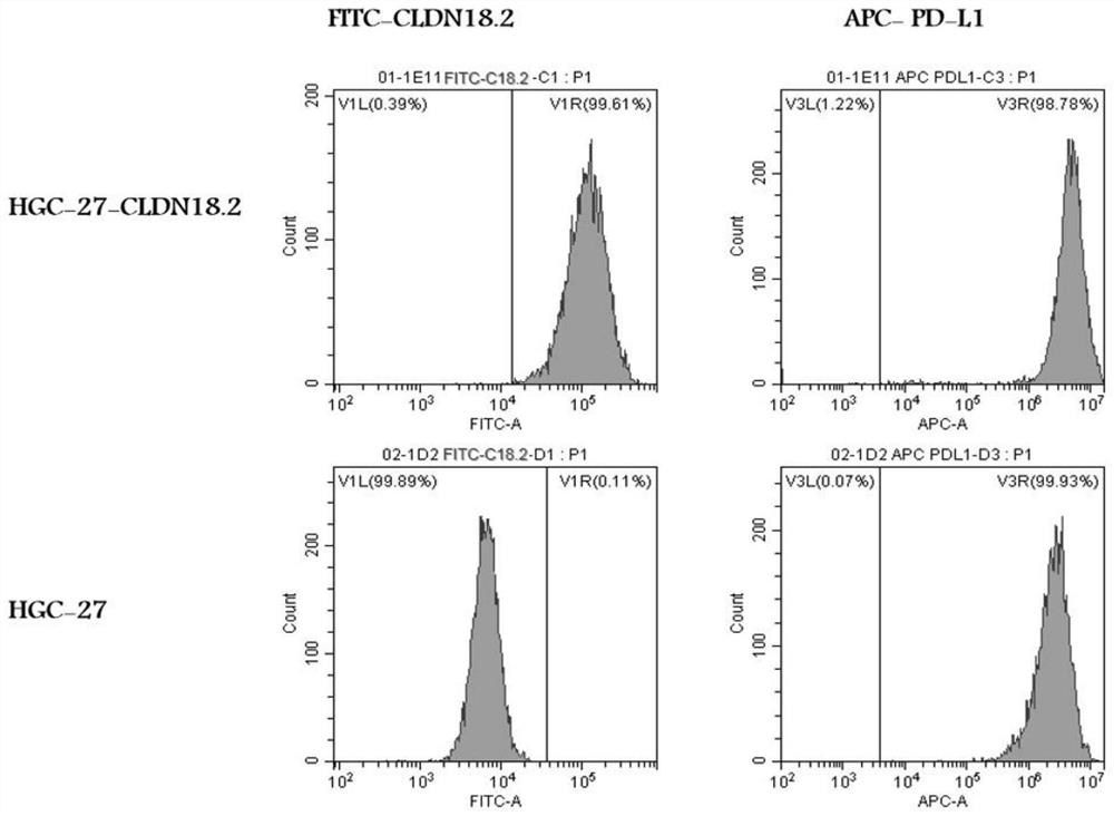
[0066] The virus supernatant was collected at 48h and 72h, and concentrated by ultracentrifugation (Merck Millipore); the concentrated virus could be used to infect HGC-27, and finally the HGC-27 cell line overexpressing CLDN18.2 was obtained and named as HGC-27- The detection results of CLDN18.2 and HGC-27-CLDN18.2 cells expressing CLDN18.2 are as follows figure 2 shown.
[0067] S200: Isolation of peripheral blood PBMC and expansion of T cells
[0068] Mononuclear cells were isolated from donor peripheral blood, subjected to density gradient centrifugation using ficol, and enriched for T cells using a T cell sorting kit (CD3 MicroBeads, human-lyophilized, 130-097-043) using conjugated anti-CD3 /anti-CD28 magnetic beads activate the culture and expansion of T cells;
[0069] The medium used TexMACS GMP Medium (Miltenyi Biotec, 170-076-309), containing 10% FBS, 2mM L-glutamine, 100IU/ml rhIL2, all cells were placed at 37°C, 5% CO 2 Cultured in a constant temperature incubator.
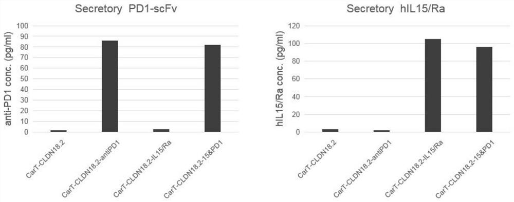
[0070] 1) The proteins in the B#~D# sequences in the structural design of the fusion protein were expressed and purified by the CHO protein expression system as a positive control standard for ELISA detection of protein secretion in 1#~4#, and the CarT culture supernatant was collected. Serum protein secretion data such as image 3 As shown, CarT-CLDN18.2-IL15/Ra, CarT-CLDN18.2-antiPD1, CarT-CLDN18.2-15&PD1 can secrete proteins normally, and have substantially the same protein secretion efficiency.
[0071] CarT was prepared by lentiviral packaging. The positive rate and phenotype results are shown in Figure 5 and Image 6. The obtained CarT proliferation is as follows Figure 4 As shown, CarT-CLDN18.2-15&PD1 has more excellent cell proliferation ability.