Cell nucleus segmentation method, system and device and cancer auxiliary analysis system and device based on pathological image

A cell nucleus and image technology, applied in the field of medical imaging, can solve the problem that the accuracy of edge segmentation needs to be improved, and achieve the effect of high segmentation

Active Publication Date: 2021-08-06
湖南医药学院
View PDF7 Cites 1 Cited by
  • Summary
  • Abstract
  • Description
  • Claims
  • Application Information

AI Technical Summary

Problems solved by technology

[0005] The present invention aims to solve the problem that the accuracy of edge segmentation needs to be improved in the process of segmenting the feature map by the current neural network

Method used


Image

  • Cell nucleus segmentation method, system and device and cancer auxiliary analysis system and device based on pathological image
  • Cell nucleus segmentation method, system and device and cancer auxiliary analysis system and device based on pathological image

Examples


specific Embodiment approach 1

[0067] This embodiment is a cell nucleus segmentation method, comprising the following steps:

[0068] 1. Collect stained images of cancer slices to construct an image set, and divide the image set into a training set and a test set.

[0069] The process collects stained images of certain cancer slices, and the stained slice images of the process are obtained by using stained slices made in actual work. Considering the workload and difficulty of collecting images and labeling, this implementation method identifies images of cervical cancer and performs corresponding model training; in fact, the stained images of slices are obtained after slicing, staining, scanning and other processes, among which The staining process can be any effective staining method. The stained image of the slice in this embodiment is an image of cervical cancer, and the 40X rendering of the stained slice is selected.

[0070] The ratio of the number of pathological slices in the training set and the te...

specific Embodiment approach 2

[0103] This embodiment is a cell nucleus segmentation system, and the system includes:

[0104] The stained slice image acquisition module is used to acquire the stained slice image and divide the image into image blocks;

[0105] The image segmentation module calls the cell nucleus segmentation network model to perform cell nucleus segmentation on the image block;

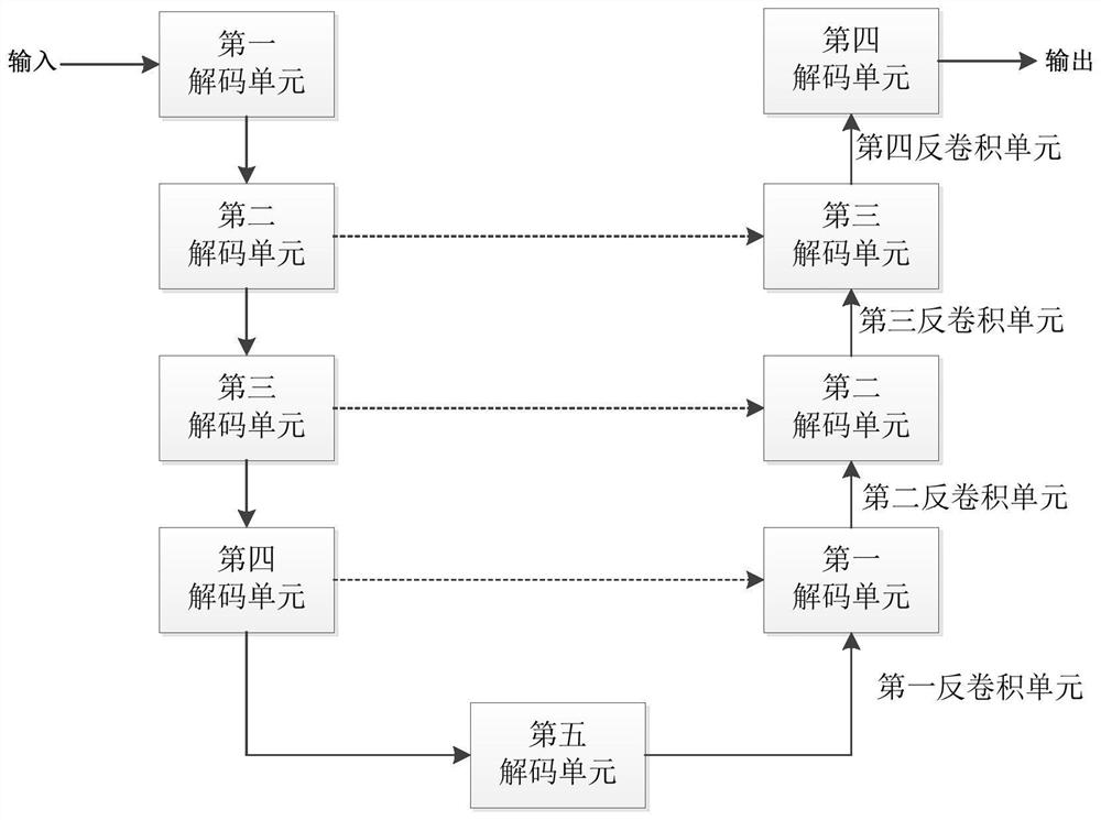
[0106] The described cell nucleus segmentation network model adopts encoder-decoder network structure, specifically as follows:

[0107] The encoder includes five coding units, namely the first coding unit to the fifth coding unit, and the image blocks are sequentially processed through the first coding unit to the fifth coding unit; wherein,

[0108] The first encoding unit includes 1 5*5 convolution, 1 BN layer, 1 activation function layer and 1 pooling layer;

[0109] The second coding unit to the fifth coding unit respectively include 3 convolution groups, 4 convolution groups, 4 convolution groups, and 3 co...

specific Embodiment approach 3

[0129] This embodiment is a cell nucleus segmentation device, and the device is used for storing and / or operating a cell nucleus segmentation system. The devices described in this embodiment include but are not limited to storage media, computers, servers, mobile devices, and the like.


PUM


Abstract

The invention discloses a cell nucleus segmentation method, system and device and a cancer auxiliary analysis system and device based on a pathological image, and belongs to the technical field of medical images. The invention aims to solve the problem that the edge segmentation accuracy needs to be improved in the process of segmenting a feature map by a neural network at present. The cell nucleus segmentation method provided by the invention comprises the following steps: aiming at a to-be-detected sample, preparing a slice and dyeing to obtain a slice dyeing image; performing image block segmentation on the section dyeing image; and then performing nucleus segmentation on an image block corresponding to the section dyeing image of the to-be-detected sample by using the nucleus segmentation network model to obtain a nucleus boundary segmentation image. According to the cancer auxiliary analysis system based on the pathological image, a cancer auxiliary analysis module is additionally arranged on the basis of cell nucleus segmentation, cancerous cells are recognized and classified according to the segmentation result of the image segmentation module based on the expert database, and therefore cancer auxiliary analysis is achieved. The method is mainly used for nucleus segmentation and cancer auxiliary analysis.

Description

technical field [0001] The invention relates to a cell nucleus segmentation method, system and cancer auxiliary analysis system, belonging to the technical field of medical imaging. Background technique [0002] With the development and maturity of deep learning technology, deep learning technology has become the mainstream technology or research direction in many application fields, and has achieved very good recognition and detection results in many fields. [0003] At present, many researchers and scholars use deep learning technology for the segmentation and identification of cancer cells, so as to assist doctors in diagnosing and analyzing cancer and reduce the workload of doctors. Some of the existing methods of using deep learning technology to identify cancer cells start with improving the staining effect, supplemented by some conventional neural networks for identification, and some start with cell morphology and improve the neural network to improve the identificat...

Claims


Application Information

Patent Timeline
IPC IPC(8): G06T7/00G06T7/11G06K9/38G06K9/62G06N3/04G06N3/08
CPCG06T7/0012G06T7/11G06N3/08G06T2207/10056G06T2207/10024G06T2207/20021G06T2207/20081G06T2207/20084G06T2207/30024G06T2207/30096G06V10/28G06N3/048G06N3/045G06F18/241
Inventor 王晓乔张在其尹辉明阳大庆唐娜萍
Owner 湖南医药学院
Why Eureka
Try Eureka