A kind of bioactive preparation based on tumor cell membrane and its preparation method and application

A bioactive, cell membrane technology, applied in the field of medicine, to achieve good biocompatibility, excellent anti-tumor effect, and improve the effect of anti-tumor effect in vivo

Active Publication Date: 2022-07-01
SHANDONG UNIV
View PDF0 Cites 0 Cited by
  • Summary
  • Abstract
  • Description
  • Claims
  • Application Information

AI Technical Summary

Problems solved by technology

So far, there is no drug that can directly target exosomal PD-L1 in clinical practice, thus presenting a challenge for the precise regulation of exosomal PD-L1

Method used


Image

  • A kind of bioactive preparation based on tumor cell membrane and its preparation method and application
  • A kind of bioactive preparation based on tumor cell membrane and its preparation method and application
  • A kind of bioactive preparation based on tumor cell membrane and its preparation method and application

Examples


preparation example Construction

[0060] In yet another specific embodiment of the present invention, a method for preparing the above-mentioned cell membrane-based bioactive preparation is provided, comprising:

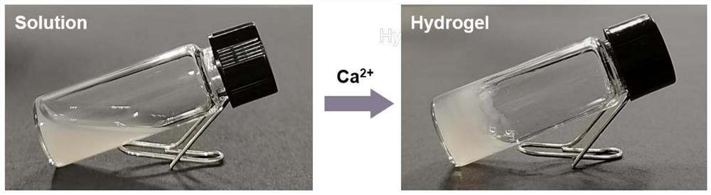
[0061] The tumor cell membrane modified by sodium alginate oxide forms a gelling factor. The CDK5 inhibitor and the exosome inhibitory drug are respectively dissolved and mixed uniformly to form a drug solution, which is added to the aqueous solution of the gelling factor and stirred to obtain a gelling solution.

[0062] In another specific embodiment of the present invention, the solvent for dissolving the CDK5 inhibitor and the exosome inhibitor is deionized water, which has a better dissolving effect, and the method of adding the drug solution is dropwise, so that the antitumor drug components are dispersed more efficiently. uniform;

[0063] In another specific embodiment of the present invention, the cell membrane vesicles are prepared by the following method:

[0064] (1) Collect mouse tumor ...

Embodiment 1

[0095] Example 1: Preparation of sodium alginate oxide-modified tumor cell membrane vesicles

[0096] 1) Preparation of tumor single cell suspension: After collecting mouse melanoma tissue, it was obtained by adding homogenization buffer containing 0.25mM sucrose, 1mM EDTA, 20mM HEPES-NaOH and protease inhibitor agent in PBS buffer.

[0097] 2) Extraction of tumor cell membranes: The collected tumor single cell suspension was subjected to probe ultrasound at 4°C to break the tumor cells to obtain broken tumor cells. Off: 2s; centrifuge the broken tumor cells at 3000g for 10min at 4°C to remove melanin for purification; centrifuge the purified broken cell suspension at 15000g for 60min at 4°C to obtain tumor cell membrane pellets; redisperse the cell membrane pellets in PBS, cryopreserved.

[0098] 3) Preparation of tumor cell membrane vesicles: The tumor cell membrane was sonicated at 4° C. for 1 min to finally obtain tumor cell membrane vesicles.

[0099]4) Preparation of ...

Embodiment 2

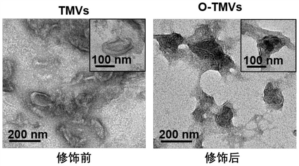
[0101] Example 2: Transmission electron microscopy (TEM) identification of nanostructures of tumor cell membrane vesicles before and after modification

[0102] Pipette 10 μL of tumor cell membrane vesicle solution before and after modification, respectively, absorb excess liquid on carbon membrane copper mesh, filter paper, dry at room temperature and observe under transmission electron microscope. The result is as figure 1 As shown, transmission electron microscopy images can confirm that the morphology of tumor cell membrane vesicles before and after modification is significantly different, with sizes of ~160 and ~200 nm, respectively, with uniform size and good dispersion.


PUM

PropertyMeasurementUnit
sizeaaaaaaaaaa

Abstract

The invention provides a biologically active preparation based on tumor cell membrane, a preparation method and application thereof, and belongs to the technical field of medicine. In the present invention, the tumor tissue of tumor-bearing mice is extracted, processed and processed to form tumor cell membrane vesicles, and after chemical modification, a gelling factor that can form a gel in situ in the tumor tissue is formed. (CDK5) inhibitor and exosome inhibitor can effectively regulate tumor cells and tumor exosomal PD-L1, thereby simultaneously releasing local and systemic immunosuppression, so it has good practical application value.

Description

technical field [0001] The invention belongs to the technical field of medicine, and in particular relates to a biologically active preparation based on tumor cell membranes and a preparation method and application thereof. Background technique [0002] The information disclosed in this Background section is only for enhancement of understanding of the general background of the invention and should not necessarily be taken as an acknowledgement or any form of suggestion that this information forms the prior art already known to a person of ordinary skill in the art. [0003] Programmed death 1 ligand (PD-L1) is generally thought to act on the surface of tumor cells, yet the failure rate of PD-L1 blockade-based immunotherapy in clinical cases remains high. Current approaches to enhance the response efficiency of PD-L1-based therapies are limited to locally modulating the tumor site, such as increasing tumor immunogenicity and remodeling the tumor's immune microenvironment, wh...

Claims


Application Information

Patent Timeline
Patent Type & Authority Patents(China)
IPC IPC(8): A61K45/06A61K47/61A61K47/69A61K9/06A61P35/00A61P37/04
CPCA61K45/06A61K9/06A61K47/61A61K47/6901A61P35/00A61P37/04
Inventor 栾玉霞李倩
Owner SHANDONG UNIV
Why Eureka
Try Eureka