
Cell image segmentation method based on deep learning

An image segmentation and deep learning technology, applied in the field of image processing, can solve the problems of insufficient precision, high complexity of cell images, and low accuracy.

Pending Publication Date: 2021-09-10
HANGZHOU DIANZI UNIV
View PDF0 Cites 0 Cited by
  • Summary
  • Abstract
  • Description
  • Claims
  • Application Information

AI Technical Summary

Problems solved by technology

[0006] However, compared with other medical images, cell images have higher complexity, and the direct use of Mask R-CNN often has problems of insufficient precision and low accuracy.

Method used


Image

  • Cell image segmentation method based on deep learning
  • Cell image segmentation method based on deep learning
  • Cell image segmentation method based on deep learning

Examples


Embodiment

[0039] In this embodiment, the specific steps of the cell image segmentation method based on deep learning are as follows:

[0040] (1) Acquire the image of the cell plate.

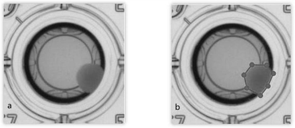
[0041] The image of the cell plate is obtained by taking an overhead view of the cell culture plate, which contains a large number of cell culture tubes arranged in an array. It is necessary to use the labelme software to label the test tube and label the cells existing in it, and the corresponding label is colony. Each image generated after annotation will generate a corresponding json file. Annotation results such as figure 2 As shown, where a is the picture before labeling; b is the picture after labeling.

[0042] (2) Establish digital image data set

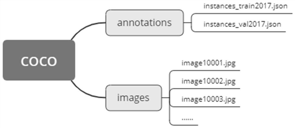
[0043] Process the json file generated after labeling, divide the digital image dataset into training set, verification set and test set, and use the labelme2coco.py file to convert it into the folder structure of the coco dataset required for Mask...


PUM


Abstract

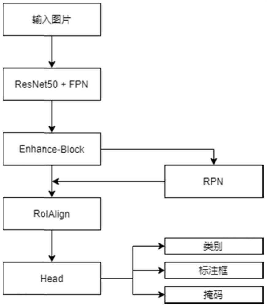
The invention discloses a cell image segmentation method based on deep learning, and belongs to the field of image processing. The method specifically comprises the following steps: segmenting cells in test tube holes by using an improved Mask R-CNN (Region-Convolutional Neural Network), obtaining cell images, establishing a digital image data set, fusing feature learning into a model establishing process, and accurately detecting and segmenting the cells in the test tube holes. A quick, low-cost and accurate automatic detection technology is realized, and the detection efficiency can be improved.

Description

technical field [0001] The invention belongs to the field of image processing, and in particular relates to a cell image segmentation method based on deep learning. Background technique [0002] In medical image processing, it is often necessary to segment the cells in the image from the background to analyze changes in their outlines and internal structures, thereby providing a basis for medical identification. Traditional cell image segmentation needs to be done manually, but when the number of cells in the image is large, the manual segmentation method often cannot meet the application requirements in terms of efficiency. Therefore, in recent years, with the continuous development of artificial intelligence, machine learning technology has been gradually introduced into cell image segmentation. This is the inevitable result of the development of both theory and technology, and has extremely promising application prospects. [0003] Machine learning is usually implemented...

Claims


Application Information

Patent Timeline
Patent Type & Authority Applications(China)
IPC IPC(8): G06T7/00G06T7/11G06N3/04G06N3/08
CPCG06T7/0012G06T7/11G06N3/08G06T2207/10056G06T2207/20081G06T2207/20084G06T2207/30024G06N3/048G06N3/045Y02T10/40
Inventor 陆剑锋缪志刚王兴伟张彬彬陈作磊俞韬
Owner HANGZHOU DIANZI UNIV
Learn More