
Cerebrovascular pathology viewing and treatment apparatus

A technology of cerebrovascular and equipment, applied in the field of cerebrovascular disease observation and treatment equipment, which can solve problems such as bleeding complications, misplacement of treatment devices, and thromboembolism in patients

Pending Publication Date: 2021-09-21
VENA MEDICAL HOLDINGS CORP
View PDF10 Cites 1 Cited by
  • Summary
  • Abstract
  • Description
  • Claims
  • Application Information

AI Technical Summary

Problems solved by technology

Furthermore, such indirect visualization using radiofluorescence imaging equipment in difficult anatomical situations may lead to misplacement of the treatment device, which may lead to thromboembolic and hemorrhagic complications in the patient in the case of cerebrovascular lesions

Method used


Image

  • Cerebrovascular pathology viewing and treatment apparatus
  • Cerebrovascular pathology viewing and treatment apparatus
  • Cerebrovascular pathology viewing and treatment apparatus

Examples


Embodiment approach

[0090]

[0091]

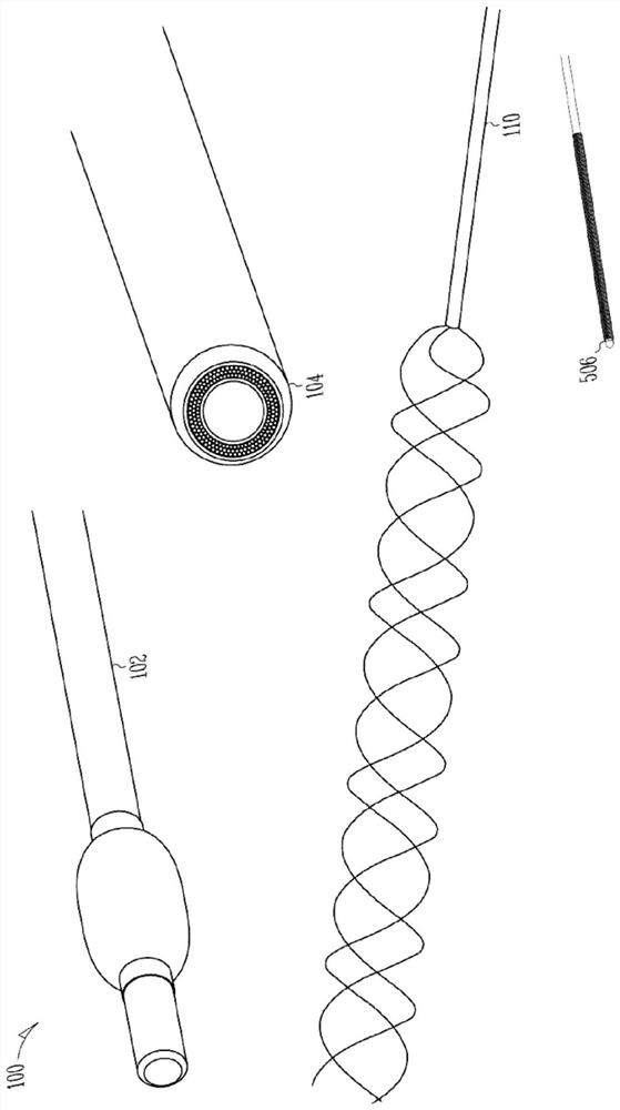
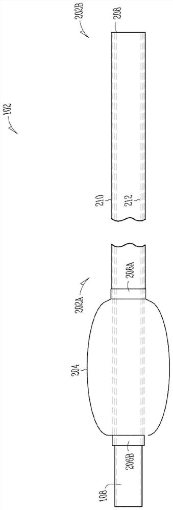
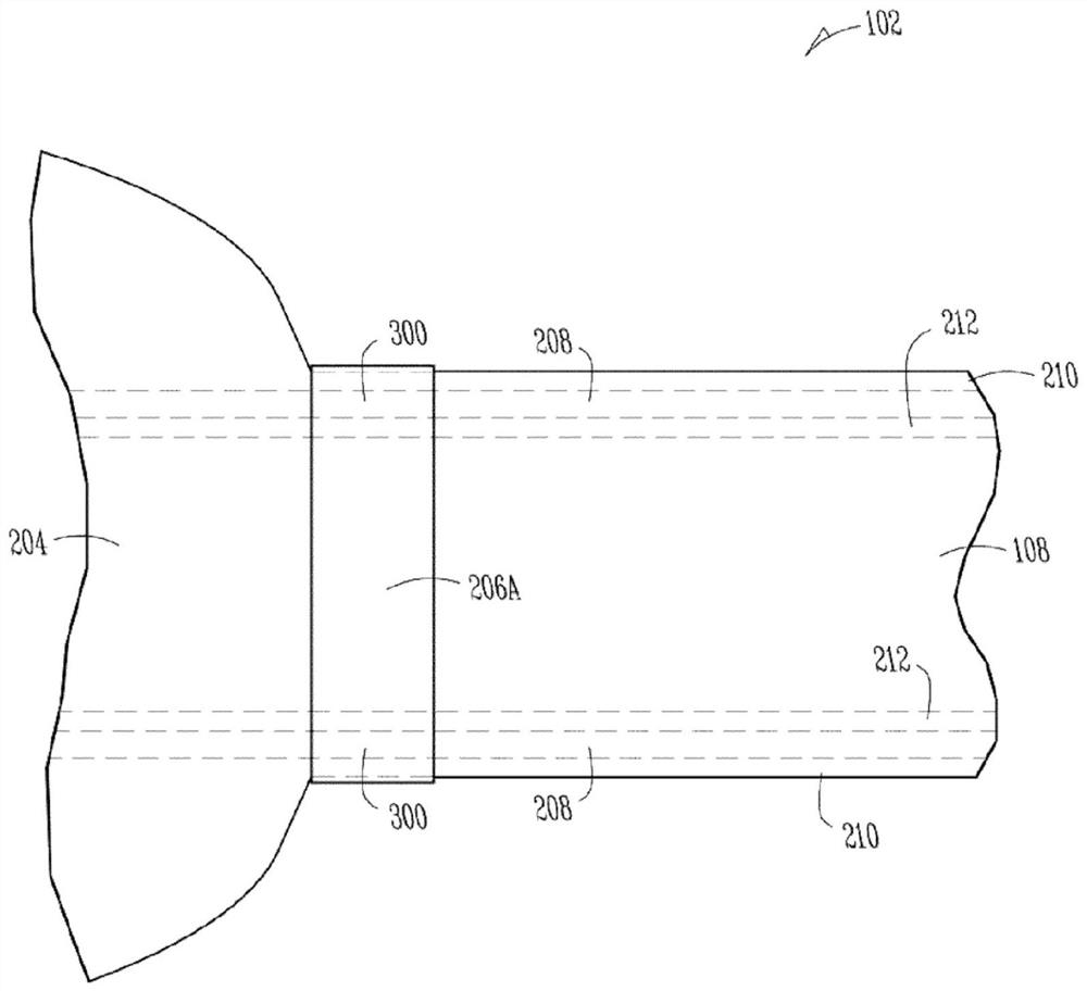
[0092] Inner layer 922 may include PTFE or other lining, such as may help provide desired lubricity to the inner walls of working chamber 108 . One or more portions of the liner or other inner layer 922 may be stretched, for example, between 50% and 200%, inclusive, or another desired stretch amount, to Provides additional bending flexibility in the area. Intermediate layer 924 may comprise a metallic hypotube, such as may comprise Nitinol (e.g., Nitinol) or other structures, such as may help provide structural rigidity—including under balloon 204 or elsewhere during inflation, To help maintain the patency of the inflation lumen 207 or the working lumen 108 , including maintaining the patency of the inflation lumen 207 or the working lumen 108 during inflation of the balloon 204 . One or more portions of the metal tube or other intermediate layer 924 may be stretched, for example, between 50% and 200%, inclusive, or another desired stretch amount, to ...


PUM

PropertyMeasurementUnit
Outer diameteraaaaaaaaaa

Abstract

Cerebrovascular treatment at an intracranial location beyond the petrous segment of the carotid artery can be challenging due to blood vessel size and tortuosity. First pass cerebrovascular thrombectomy success rate under only fluoroscopic guidance can be low (e.g., 25.1%) but an angioscope can help improve efficacy. A sheath catheter can be advanced toward the cerebrovascular pathology. Its distal balloon can be inflated. An angioscope can be inserted via its working lumen for viewing. The sheath catheter can have a stepped-down lateral profile and can extend the working channel a distance beyond the balloon. A dual concentric lumen structure can include an inner body and an outer body, defining an inflation lumen therebetween, with one or more portions of one or more layers stretched or cut or both, such as to provide bending flexibility. Reflow techniques can be used to help bond layers together.

Description

[0001] priority claim [0002] This application claims the benefit of priority to U.S. Provisional Patent Application Serial No. 62 / 790,330, filed January 9, 2019, which is incorporated herein by reference in its entirety. technical field [0003] This document relates generally but not limited to intravascular imaging and therapy, and more particularly but not limited to cerebrovascular disease observation and treatment. Background technique [0004] Cerebrovascular lesions may include, among others, thrombi (blood clots) or aneurysms (localized enlargement or weakening of blood vessels). Cerebral blood clots can be associated with ischemic strokes, which in turn cause damage to brain tissue. Cerebrovascular aneurysms may be associated with a risk of rupture and hemorrhagic stroke, which in turn can cause damage to brain tissue. [0005] Radiofluorescence imaging of imaging device locations from outside the patient can be used to guide endovascular devices within the pati...

Claims


Application Information

Patent Timeline
IPC IPC(8): A61M25/10A61B1/00A61B1/313A61B1/07A61B17/12A61B17/22A61B90/00
CPCA61B1/313A61B1/07A61B1/00167A61B1/00082A61B2017/1205A61B17/12113A61B2017/22068A61B2017/22069A61B2017/003A61B2017/00309A61B2090/306A61B2017/22044A61B2017/22094A61B17/221A61B2017/22034A61B17/22A61B2217/005A61B90/361A61M2025/0681A61B1/3137A61B2017/22079A61B1/018A61B1/0055A61F2002/9528
Inventor 菲利普·库伯迈克尔·菲利普斯
Owner VENA MEDICAL HOLDINGS CORP
Learn More