Construction method of biosensing system for detecting physiological and pathological parameters of organ chip

An organ chip and biosensing technology, which is applied in the field of biosensing system construction, can solve problems such as the inability to realize real-time dynamic monitoring, limited operator operation technology, and long detection time, so as to reduce research costs and experiment costs , the effect of reducing the experimental error

Pending Publication Date: 2021-09-28
EAST CHINA NORMAL UNIV
View PDF7 Cites 2 Cited by
  • Summary
  • Abstract
  • Description
  • Claims
  • Application Information

AI Technical Summary

Problems solved by technology

[0003] When conventional blood vessel chips are used in the research of developmental biology and disease pathology, immunofluorescence staining is usually used to observe the chip with the help of fluorescent dyes and optical imaging methods. This method is complicated to operate and is limited by the operator's operating skills , the detection takes a long time and the cost is high. It can only analyze specific parameters and cannot realize real-time dynamic monitoring.

Method used


Image

  • Construction method of biosensing system for detecting physiological and pathological parameters of organ chip
  • Construction method of biosensing system for detecting physiological and pathological parameters of organ chip
  • Construction method of biosensing system for detecting physiological and pathological parameters of organ chip

Examples


Embodiment 1

[0096] (1) Mix the polydimethylsiloxane main agent and the curing agent evenly at a mass ratio of 10:1, and after removing air bubbles in vacuum, cast it on the pre-designed blood vessel chip motherboard, and after curing for two hours, obtain The PDMS template is separated from the master plate; the main agent is Sylgard 184 polymer, and the curing agent is Sylgard 184 curing agent.

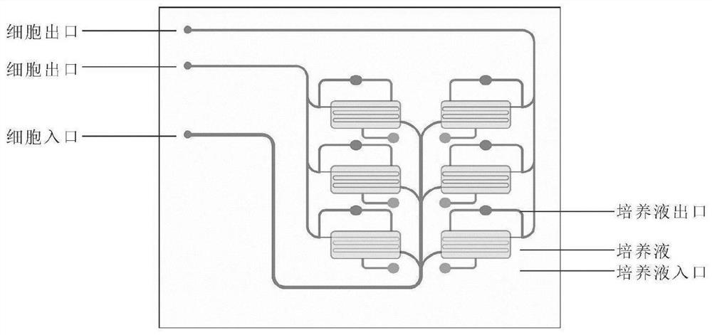
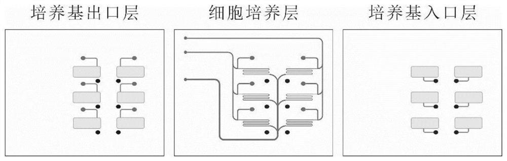
[0097] (2) Holes were punched at the inlet and outlet of the PDMS template obtained in step (1), wherein the medium outlet and inlet layers each had six 500 μm round holes, and the cell culture had three 500 μm round holes.

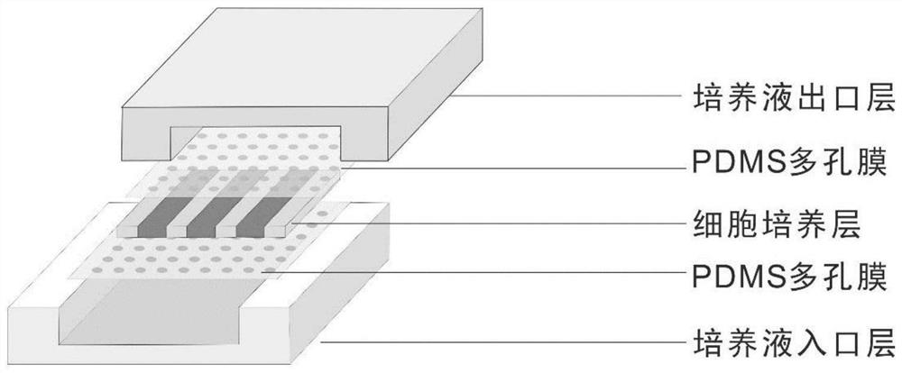
[0098] (3) The three PDMS templates prepared in step (1) were bonded layer by layer through a plasma cleaner to obtain a blood vessel chip model. The bonding conditions were: RF power 600w, time 40s, oxygen flow rate 200mL / min.

[0099] (4) will 2×10 6 Human umbilical vein endothelial cells with 2 x 10 6 Fibroblasts were mixed in a 10 mg / mL matrigel solution, and the matrig...

Embodiment 2

[0113] (1) Mix the polydimethylsiloxane main agent and the curing agent evenly at a mass ratio of 10:1, and after removing air bubbles in vacuum, cast it on the pre-designed blood vessel chip motherboard, and after curing for two hours, obtain The PDMS template is separated from the master plate; the main agent is Sylgard 184 polymer, and the curing agent is Sylgard 184 curing agent.

[0114] (2) Perforate the inlet and outlet of the PDMS template obtained in step (1), wherein the media outlet and inlet layers each have six 500 μm round holes, and the cell culture layer has three 500 μm round holes.

[0115] (3) The three PDMS templates prepared in step (1) were bonded layer by layer through a plasma cleaner to obtain a blood vessel chip model. The bonding conditions were: RF power 600w, time 40s, oxygen flow rate 200mL / min.

[0116] (4) will 2×10 6 Human umbilical endothelial cells with 2 x 10 6 Cardiomyocytes derived from personal induced pluripotent stem cells were mixed ...


PUM

PropertyMeasurementUnit
diameteraaaaaaaaaa
diameteraaaaaaaaaa
concentrationaaaaaaaaaa

Abstract

The invention discloses a construction method of a biosensing system for detecting the physiological and pathological parameters of an organ chip. The method comprises the following steps of injecting a cell mixed solution into a pre-designed chip model, and continuously culturing to form the organ chip; and then connecting an electrochemical sensing chip provided with a micro-flow control pipeline to an outlet of the organ chip for specifically monitoring the oxygen content, ATP, pH, ROS and cholesterol in metabolites of the organ chip. An organ chip analysis system is simple and convenient to construct, is wide in application range and diversified in analysis parameters; the organ chip is combined with an electrochemical sensor, so that the growth micro-environment of a three-dimensional in-vitro blood vessel / heart model can be dynamically monitored in real time. According to the electrochemical biosensing system for monitoring the physiological and pathological indexes of the organ chip, the model culture condition can be adjusted in real time, so that the model culture condition better conforms to the real living body condition, and the electrochemical biosensing system can be used for exploring the physiological state change in the disease process and also can be used for monitoring the response of the organ chip to drugs on line.

Description

technical field [0001] The invention belongs to the technical field of 3D in vitro culture and electrochemical analysis of cells, and relates to a construction method of a biosensing system for detecting physiological and pathological parameters of an organ chip, which integrates an organ chip and a plurality of electrochemical biosensors. Background technique [0002] Organ chip is a 3D in vitro tissue model that more realistically reflects the human body. During the construction of organ chip, corresponding human cell lines are cultivated in the chip model to simulate the structure and functional units of real human tissues or organs. As an emerging 3D in vitro disease model, organ chips make up for the shortcomings of existing 2D models and animal models, and have great application potential in biological development, drug development and precision medicine. [0003] When conventional blood vessel chips are used in the research of developmental biology and disease patholo...

Claims


Application Information

Patent Timeline
Patent Type & Authority Applications(China)
IPC IPC(8): G01N27/327
CPCG01N27/3278
Inventor 裴昊余紫荆李丽
Owner EAST CHINA NORMAL UNIV
Why Eureka
Try Eureka