Embryo development potential prediction method and system, equipment and storage medium

A technology of embryo development and prediction method, which is applied in the field of medical artificial intelligence, can solve problems such as inability to use, inability to fully mine embryo image features, inability to model single-focus embryo time-lapse video timing, etc., to achieve reliable prediction results

Active Publication Date: 2021-10-01
THE FIRST AFFILIATED HOSPITAL OF SUN YAT SEN UNIV
View PDF4 Cites 4 Cited by
  • Summary
  • Abstract
  • Description
  • Claims
  • Application Information

AI Technical Summary

Problems solved by technology

[0003] With the development of assisted reproductive technology, the proportion of Chinese couples who choose to obtain newborns through assisted reproductive treatment is increasing year by year. Among them, in vitro fertilization has become a key technology for the treatment of infertility. In vitro fertilization technology uses artificial methods to make eggs and sperm In vitro fertilization, and early embryo development, and then transplanted into the mother's uterus for pregnancy and development, after maturity, delivery in a normal way. However, in terms of clinical pregnancy rate prediction after embryo transfer, the existing methods not only require doctors to monitor the morphological changes of embryos A large number of labels, using a small number of artificially produced morphological parameters to directly perform data-label mapping learning, and only use a small amount of clinical data of patients and a small number of artificially formulated embryo dynamic parameters for single-focus embryo time-lapse (time-lapse photography) video The analysis cannot fully exploit the image features of embryonic development in the single-focus embryo time-lapse video, nor can it capture the temporal and spatial characteristics of the single-focus embryo time-lapse video, resulting in a serious lack of interpretability for the pregnancy rate prediction results, and the experiment Results are highly dependent on the size and balance of the data
[0004] In addition, due to the limited survival rate of embryos (fertilized eggs) in vitro culture, it is generally required to culture multiple embryos in each cycle of assisted fertility treatment, and only embryos that have developed to the blastocyst stage will be considered for transplantation. Whether the embryos form blastocysts is a key step in in vitro fertilization and an important basis for subsequent embryo selection and transfer. However, the existing technology can only analyze the complete single-focus embryo time-lapse video and detect the cultured embryos, and cannot predict embryos in real time. The probability of forming blastocysts, so that doctors cannot find embryos with a low probability of forming blastocysts in time, and stop the cultivation of embryos with low developmental potential at an early stage
[0005] At the same time, in terms of predicting whether the blastocyst is euploid, there are existing methods for predicting whether the embryo is euploid by observing a single picture, such as: using the pre-trained ResNet-152 model to predict whether the blastocyst is euploid , can not perform sequential modeling on single-focus segment embryo time-lapse video, resulting in low euploid prediction accuracy and poor interpretability, and cannot be used in actual clinical scenarios

Method used


Image

  • Embryo development potential prediction method and system, equipment and storage medium
  • Embryo development potential prediction method and system, equipment and storage medium
  • Embryo development potential prediction method and system, equipment and storage medium

Examples


Embodiment Construction

[0056] The embodiment of the present invention will be explained in detail below in conjunction with the accompanying drawings. The examples given are only for the purpose of illustration, and cannot be interpreted as limiting the present invention. The accompanying drawings are only for reference and description, and do not constitute the scope of patent protection of the present invention. limitations, since many changes may be made in the invention without departing from the spirit and scope of the invention.

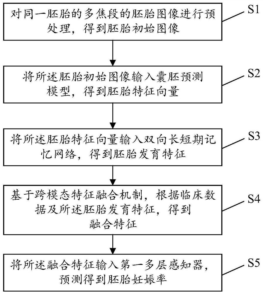
[0057] The existing methods for predicting the potential of embryonic development mainly analyze single-focus embryo time-lapse videos, which cannot make full use of multi-focus information, and cannot capture the characteristics of time-lapse videos in time and space, resulting in inability to efficiently and accurately predict embryos. For questions about pregnancy rates, see figure 1 , figure 1 It is a schematic flow chart of a method for predicting embryonic dev...


PUM


Abstract

The invention relates to the technical field of medical artificial intelligence, in particular to an embryo development potential prediction method and system, equipment and a storage medium. The method comprises the steps: inputting an embryo initial image into a blastocyst prediction model, and obtaining an embryo feature vector; inputting the embryo feature vector into a bidirectional long-short-term memory network to obtain embryo development features; based on a cross-modal feature fusion mechanism, obtaining fusion features according to clinical data and the embryonic development features; and inputting the fusion features into a first multi-layer sensor, and predicting to obtain the embryo pregnancy rate. According to the invention, multi-focal-segment embryo videos shot in the early stage are analyzed, and the fusion features with the space-time characteristic is obtained by utilizing a multi-focal-segment selection model and a time transfer model, so that the pregnancy rate of the embryo cultured in vitro is predicted in real time, and the prediction accuracy is improved; meanwhile, by predicting the blastocyst forming probability and the euploid probability, doctors are assisted in early embryo screening, and therefore the labor cost is reduced.

Description

technical field [0001] The present invention relates to the technical field of medical artificial intelligence, in particular to a method, system, device and storage medium for predicting embryonic developmental potential based on multi-focus segment time-lapse video. Background technique [0002] In recent years, due to reasons such as delayed childbearing age and high life pressure, the infertility rate of couples of childbearing age in China has climbed to 12%-15%, and the number of patients has exceeded 50 million. the third leading disease. [0003] With the development of assisted reproductive technology, the proportion of Chinese couples who choose to obtain newborns through assisted reproductive treatment is increasing year by year. Among them, in vitro fertilization has become a key technology for the treatment of infertility. In vitro fertilization technology uses artificial methods to make eggs and sperm In vitro fertilization, and early embryo development, and t...

Claims


Application Information

Patent Timeline
Patent Type & Authority Applications(China)
IPC IPC(8): G06T7/00G06K9/62G06N3/04
CPCG06T7/0012G06T2207/30044G06N3/044G06F18/2415G06F18/253Y02A90/10
Inventor 麦庆云李冠彬高峰周灿权颜鹏翔陈方莹谢翔丁晨晖徐艳文
Owner THE FIRST AFFILIATED HOSPITAL OF SUN YAT SEN UNIV
Why Eureka
Try Eureka