Navigation marker for magnetic resonance and CT

A magnetic resonance and marker technology, applied in the field of navigation markers, can solve the problems of long scanning time, expensive second magnetic resonance equipment, low density, etc., and achieve the effects of avoiding accidental drop, remarkable navigation effect and simple structure

Pending Publication Date: 2021-10-08
THE AFFILIATED HOSPITAL OF SOUTHWEST MEDICAL UNIV
View PDF0 Cites 0 Cited by
  • Summary
  • Abstract
  • Description
  • Claims
  • Application Information

AI Technical Summary

Problems solved by technology

Conventional navigation markers show low signal in MRI T2 sequences and MRI enhanced sequences, and are not easy to be identified. Some navigation markers on the scalp are discarded and wasted due to unclear display, which reduces the accuracy of neuronavigation to a certain extent. Spend
The first MRI scan time is relatively long, and the second MRI equipment is expensive. Primary hospitals often do not have MRI equipment. Navigation based on CT data can replace MRI navigation in units without MRI. Conventional CT navigation markers contain metal. , metal artifacts will be formed after CT scanning, which will interfere with the display of brain structures and lesions
Conventional magnetic resonance navigation markers are used for CT navigation markers because of their lower density, more unclear display, and less easy to identify

Method used


Image

  • Navigation marker for magnetic resonance and CT

Examples


Embodiment 1

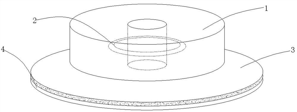
[0027] The main structure of this embodiment, such as figure 1 As shown, it includes a main body 1 with a through hole in the middle. A completely closed circular tube 2 is embedded inside the main body 1. The circular tube 2 is filled with vegetable oil. The outer edge of the bottom of the main body 1 is provided with a The adhesive part 3 and the main body 1 are made of acrylonitrile-styrene-butadiene copolymer.

[0028] The specific implementation process is that after the patient shaves his head before the operation, a magnetic resonance and CT dual-purpose navigation marker is pasted on the scalp, even if the adhesive part 3 at the bottom of the main body 1 sticks to the surface of the patient's scalp, and then the magnetic resonance or CT scan is performed. The DICOM data is imported into the navigation system, and the three-dimensional navigation and positioning of the brain lesion can be performed by inserting the navigation probe into the through hole in the middle of...

Embodiment 2

[0030] In this embodiment, on the basis of the above embodiments, an anti-adhesive release paper 4 is further added, such as figure 1 As shown, it also includes an anti-adhesive release paper 4 that matches the shape of the adhesive part 3 , and one side of the anti-adhesive release paper 4 is a smooth surface closely bonded to the adhesive part 3 . The setting of the anti-adhesive release paper 4 is to prevent the adhesive part 3 from being polluted by sundries when it is not in use. For the patient's scalp, just place the main body 1 on the surface of the patient's scalp. Other parts of this embodiment are the same as those of the foregoing embodiment, and will not be repeated here.

Embodiment 3

[0032] This embodiment further defines the structure of the main body 1 on the basis of the above embodiments, such as figure 1 As shown, the main body 1 is cylindrical. Here, the shape of the main body 1 is preferably cylindrical, and other shapes of the main body 1 can also be selected without affecting the navigation marks. Other parts of this embodiment are the same as those of the foregoing embodiment, and will not be repeated here.


PUM

PropertyMeasurementUnit
heightaaaaaaaaaa
diameteraaaaaaaaaa

Abstract

The invention discloses a navigation marker for magnetic resonance and CT (Computed Tomography), which comprises a main body with a through hole in the middle, a completely closed annular circular tube is embedded in the main body, vegetable oil is filled in the annular circular tube, a sticking part extending outwards is arranged on the outer edge of the bottom of the main body, and the main body is made of acrylonitrile-styrene-butadiene copolymer. The navigation marker provided by the invention can be used as an MRI and CT navigation marker at the same time, the annular circular tube filled with the vegetable oil is arranged in the main body, the vegetable oil is high in signal and easy to recognize in a magnetic resonance T2 sequence and an enhanced sequence at the same time, and in addition, the main body is made of the acrylonitrile-styrene-butadiene copolymer, and is high in density and easy to recognize under CT scanning; the sticking part is additionally arranged, so that the navigation marker on the head of a patient is prevented from accidentally falling off after sleeping; and the navigation marker is simple in structure and convenient to use, provides great convenience for three-dimensional navigation and positioning of the patients with a craniocerebral lesion, and is suitable for wide popularization and application.

Description

technical field [0001] The invention relates to the technical field of medical devices, in particular to a navigation marker that satisfies the dual functions of magnetic resonance and CT. Background technique [0002] For neurosurgeons, the preoperative and intraoperative precise positioning and projection, minimally invasive, brain protection and patient prognosis of minimal brain lesions, deep brain lesions, brainstem and brain functional area lesions are very important. [0003] Taking magnetic resonance navigation as an example, after the preoperative patient shaves his head, a conventional navigation marker is pasted on the scalp, and then the magnetic resonance scan is performed. After the scan, the magnetic resonance DICOM data is imported into the navigation system to perform three-dimensional navigation and positioning of brain lesions. Conventional navigation markers show low signal in MRI T2 sequences and MRI enhanced sequences, and are not easy to be identified....

Claims


Application Information

Patent Timeline
Patent Type & Authority Applications(China)
IPC IPC(8): A61B90/00
CPCA61B90/39A61B2090/3954A61B2090/3966
Inventor 钟萍李慎杰狄滨茹杨韬孙树杰王科杨述根陈光祥兰永树陈果
Owner THE AFFILIATED HOSPITAL OF SOUTHWEST MEDICAL UNIV
Why Eureka
Try Eureka