Focus high-definition image reconstruction method and device, equipment and storage medium

A technology of high-definition images and lesions, applied in the field of image processing, can solve the problems of reducing machine usage efficiency, occupying CT equipment, increasing the workload of doctors or technicians, and achieving the effect of improving efficiency and reducing workload

Pending Publication Date: 2021-10-22
PEKING UNIV SHENZHEN HOSPITAL
View PDF7 Cites 0 Cited by
  • Summary
  • Abstract
  • Description
  • Claims
  • Application Information

AI Technical Summary

Problems solved by technology

At present, for the image reconstruction of small lesions, doctors or technicians usually need to perform cumbersome operations on CT equipment, and then send them to the workstation after completion. This image reconstruction method is inefficient and increases the workload of doctors or technicians. , at the same time, occupying CT equipment, reducing the efficiency of machine use

Method used


Image

  • Focus high-definition image reconstruction method and device, equipment and storage medium
  • Focus high-definition image reconstruction method and device, equipment and storage medium
  • Focus high-definition image reconstruction method and device, equipment and storage medium

Examples


Embodiment Construction

[0051] Embodiments of the present invention are described in detail below, examples of which are shown in the drawings, wherein the same or similar reference numerals designate the same or similar elements or elements having the same or similar functions throughout. The embodiments described below by referring to the figures are exemplary only for explaining the present invention and should not be construed as limiting the present invention.

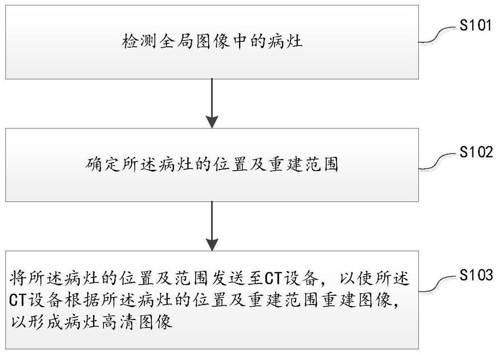
[0052] refer to figure 1 as shown, figure 1 A flowchart of an embodiment of a method for reconstructing a high-definition image of a lesion provided by an embodiment of the present invention is shown. For ease of description, only parts related to the embodiment of the present invention are shown. The method for reconstructing a high-definition image of a lesion can be performed by a CT workstation, and specifically includes: a method for reconstructing a high-definition image of a lesion, including:

[0053] S101. Detect a lesion in t...


PUM


Abstract

The invention discloses a lung nodule focus detection and automatic high-definition image reconstruction method and device, equipment, a storage medium and a computer, and the method comprises the steps of detecting a focus in a global image based on an AI technology; determining the spatial position of the focus, and determining the Z-axis reconstruction range, the image coordinate center and the FOV size of the high-definition image according to the size of the focus; and sending the position and the reconstruction range of the lesion to CT equipment, so that the CT equipment reconstructs an image according to the position and the reconstruction range of the lesion to form a lesion high-definition image. According to the focus high-definition image reconstruction method, device and equipment and the storage medium provided by the embodiment of the invention, automatic reconstruction of a small focus position VR three-dimensional image can be realized, tedious manual operation on CT equipment is not needed, the efficiency is improved, the workload of doctors or technicians is reduced, meanwhile, the CT equipment is not occupied, and the machine use efficiency is improved.

Description

technical field [0001] The present invention relates to the technical field of image processing, in particular to a method, device, equipment and storage medium for automatic reconstruction of high-definition images of lesions. Background technique [0002] CT (Computed Tomography), that is, computerized tomography, uses a precisely collimated X-ray beam to rotate around the human body synchronously with a highly sensitive detector. After the detector collects the X-ray attenuation information of the human body, it uses a special reconstruction algorithm A series of cross-sectional images are reconstructed. CT has the characteristics of fast scanning time and clear images, and can be used for the examination of various diseases. [0003] In the related art, after the original CT image obtained by CT scanning, it is generally necessary to post-process the original CT image to create a VR three-dimensional image, which is convenient for doctors to determine and observe the lo...

Claims


Application Information

Patent Timeline
Patent Type & Authority Applications(China)
IPC IPC(8): G06T5/00G06T7/00G06T7/11
CPCG06T5/001G06T7/0012G06T7/11G06T2207/10081G06T2207/30096
Inventor 袁知东
Owner PEKING UNIV SHENZHEN HOSPITAL
Why Eureka
Try Eureka