Signal noise reduction method for low-frequency ultrasonic thoracic cavity imaging

A thoracic cavity and signal technology, applied in the directions of ultrasound/sonic/infrasonic image/data processing, ultrasound/sonic/infrasonic diagnosis, ultrasound/sonic/infrasonic Permian technology, etc., which can solve the problem of few research results and the effect of imaging and other problems, to achieve the effect of improving the smoothness index, reducing the root mean square error, and having a good application prospect

Pending Publication Date: 2021-10-29
SHENYANG POLYTECHNIC UNIV
View PDF0 Cites 0 Cited by
  • Summary
  • Abstract
  • Description
  • Claims
  • Application Information

AI Technical Summary

Benefits of technology

The technical effects are achieved through this new approach called COSMICOMEL (Composite Microsystem Computational Intelligence), which helps identify signals from lung tissue accurately during medical procedures such as CT scans or MRI scanning. It also improves image quality with reduced background clutter caused by scattered particles on the surface being imaged.

Problems solved by technology

The technical problem addressed in this patented text relates to low frequency sonography (LFUS) images that contain significant amounts of background noise caused from different structures within the body such as heart muscle walls and ribs.

Method used


Image

  • Signal noise reduction method for low-frequency ultrasonic thoracic cavity imaging
  • Signal noise reduction method for low-frequency ultrasonic thoracic cavity imaging
  • Signal noise reduction method for low-frequency ultrasonic thoracic cavity imaging

Examples


Embodiment

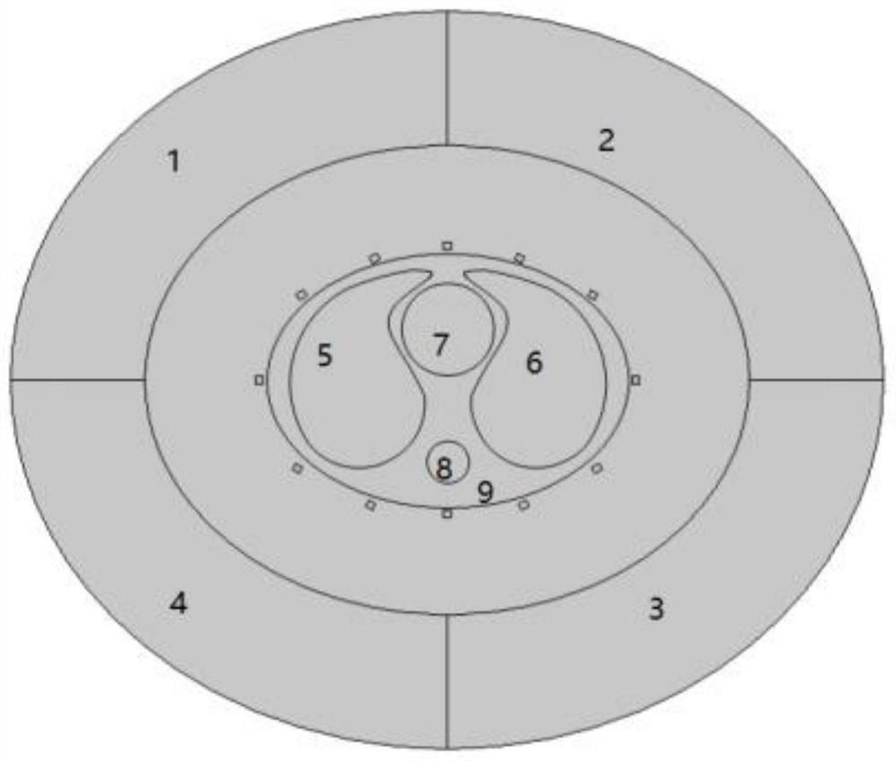
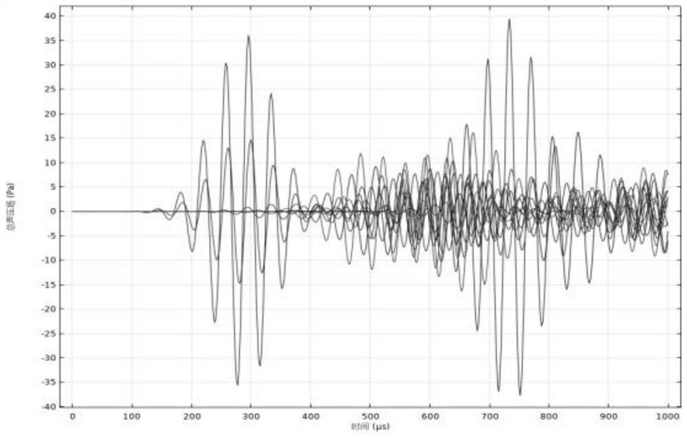
[0039]The present invention uses the db wavelet family, the sym wavelet family and the coif wavelet family to perform 1 to 10 layers of decomposition and noise reduction processing on the chest signal after noise addition, and compares the improvement of the signal-to-noise ratio after signal noise reduction to obtain the optimal mother wavelet and Optimal number of decomposition layers. In this experiment, ultrasonic transducer 1 is used to transmit ultrasonic waves at a frequency of 40kHZ, and ultrasonic transducer 2 is used to receive chest signals. Since white noise is a random number sequence, the evaluation index will fluctuate up and down every time it is obtained. Here, the average value of 50 times is taken for the signal-to-noise ratio of the chest signal after noise addition and noise reduction. The experimental data are shown in Tables 1 to 3.

[0040] Table 1 The signal-to-noise ratio (dB) of noise-added thoracic signal and db wavelet family noise reduction

[00...


PUM


Abstract

The invention discloses a signal noise reduction method for low-frequency ultrasonic thoracic cavity imaging, which comprises the following steps: modeling through COMSOL, establishing a left lung, a right lung, a heart, a spine and a skeletal muscle in the model, uniformly placing 12 ultrasonic transducers around the outside of the skeletal muscle, and setting the material as PZT-5H; adding physical fields of 'pressure acoustics, transient state', 'solid mechanics', 'static electricity ', 'sound-structure boundary' and'piezoelectric effect' into COMSOL, and building a two-dimensional thoracic cavity model; distributing the 12 piezoelectric ultrasonic transducers which are integrated in a receiving and transmitting mode evenly outside the thoracic cavity in an oval arrangement mode, the interval between every two ultrasonic transducers being 30 degrees, coupling the ultrasonic transducers with the thoracic cavity through water, and setting the perfect matching layer on the outermost portion of the simulation area. A model is established through COMSOL, and a signal noise reduction method in the field of low-frequency ultrasonic thoracic cavity imaging is creatively invented according to a one-transmitting and 11-receiving transmitting-receiving mode. According to the technology, the signal-to-noise ratio of the chest signals after noise adding can be improved, and the good noise reduction effect is achieved.

Description


Claims


Application Information

Patent Timeline
Owner SHENYANG POLYTECHNIC UNIV
Why Eureka
Try Eureka