Eureka AIR delivers breakthrough ideas for toughest innovation challenges, trusted by R&D personnel around the world.

Built-in examination and diagnosis device for anorectal department

A diagnostic device, anorectal technology, applied in the directions of diagnosis, endoscopy, proctoscope, etc., can solve the problems of poor lubricity of the device, easy adhesion of liquid to the camera lens, affecting the clarity of the lens, etc., to improve work efficiency, The effect of improving lubricity

Inactive Publication Date: 2021-11-05
THE SECOND HOSPITAL OF HEBEI MEDICAL UNIV
View PDF1 Cites 1 Cited by
  • Summary
  • Abstract
  • Description
  • Claims
  • Application Information

AI Technical Summary

Problems solved by technology

[0005] The purpose of the present invention is to solve the problems in the prior art that the camera lens is easy to adhere to the liquid, which affects the clarity of the lens and the device has poor lubricity and cannot be adjusted in the anorectum, and proposes a built-in inspection and diagnosis device for the anorectal department

Method used


Image

  • Built-in examination and diagnosis device for anorectal department
  • Built-in examination and diagnosis device for anorectal department
  • Built-in examination and diagnosis device for anorectal department

Examples


Embodiment 1

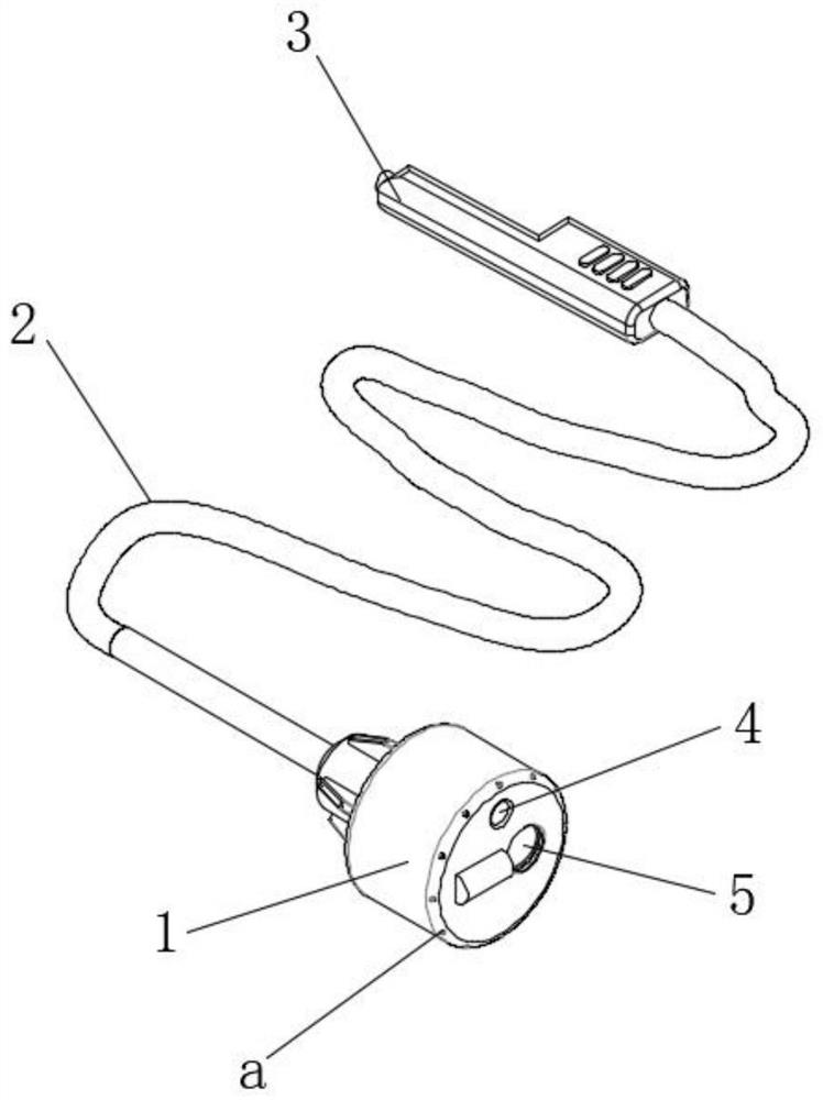
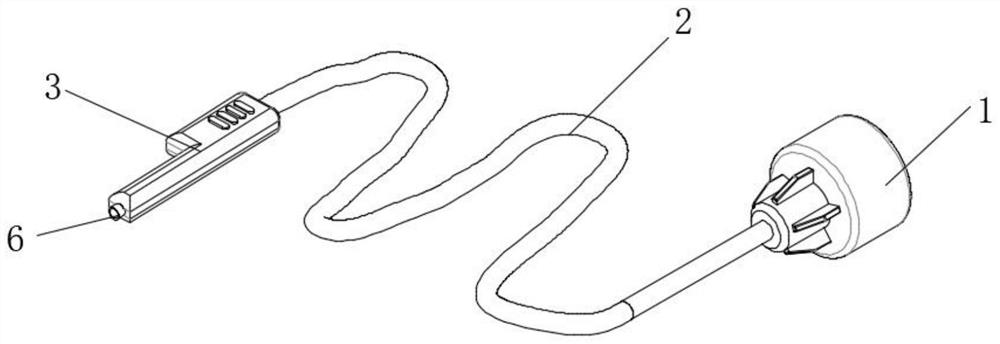
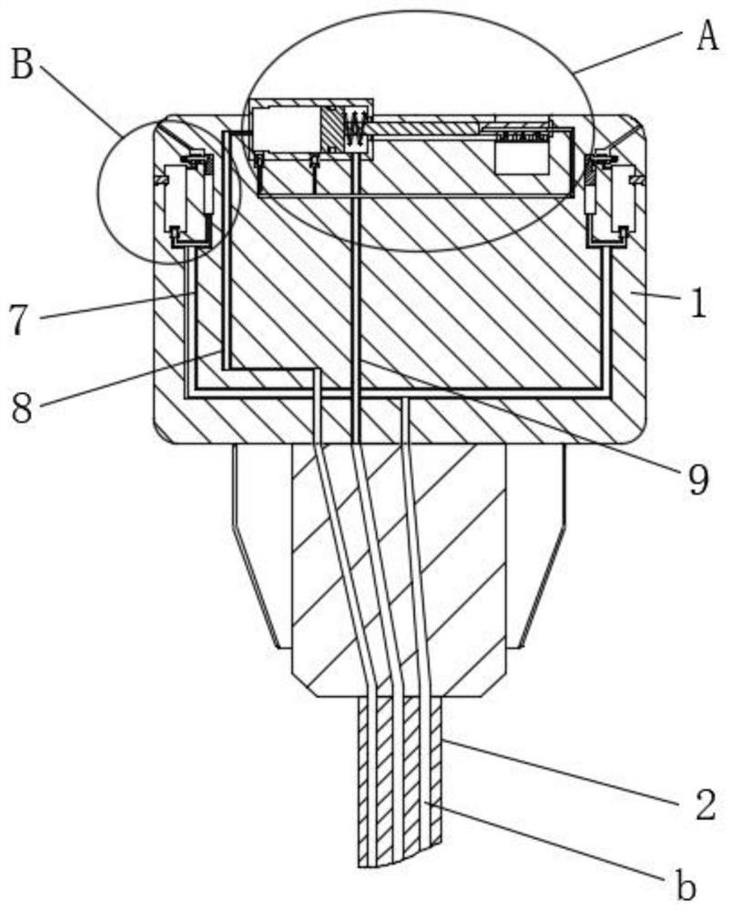
[0033] refer to Figure 1-8 , a built-in inspection and diagnosis device for anorectal department, comprising a search head 1, a resin tube 2 and a handle 3, a search light 4 and a camera 5 are arranged on one end of the search head 1, the search light 4 is located on one side of the camera 5, and the resin tube 2 is at one end It is fixedly connected with the search head 1, and the other end is fixedly connected with the handle 3. A cylinder body 10 is installed on one end of the search head 1, and the cylinder body 10 is located at one side of the camera 5. The cylinder body 10 is slidably connected with a piston 14, and the piston 14 will The inner cavity of the cylinder body 10 is divided into a left cavity and a right cavity, and one end of the piston 14 is fixedly connected with a piston rod 16, and one end of the piston rod 16 is fixedly connected with a movable plate 1601, and the movable plate 1601 is slidably connected with the search head 1 and is located outside the...

Embodiment 2

[0041] Such as Figure 1-8 As shown, this embodiment is basically the same as Embodiment 1. Preferably, several through holes are opened on the surface of the movable plate 1601 , and the through holes are located on the side of the screen pipe 18 .

[0042] In this embodiment, when the screen tube 18 is blown with air, the air flow can communicate with the outside through the through hole to form a gas circulation.

Embodiment 3

[0044] Such as Figure 1-8 As shown, this embodiment is basically the same as Embodiment 1, preferably, the screen tubes 18 and the gauze strips 17 are distributed alternately.

[0045] In this embodiment, when the screen tube 18 blows air, the air flow can push the liquid on the camera 5 to move to the gauze strips 17 on both sides and be absorbed.


PUM


Abstract

The invention discloses a built-in examination and diagnosis device, which comprises a searchlight head, a resin tube and a grab handle, wherein one end surface of the searchlight head is provided with a searchlight and a camera; the searchlight is positioned on one side of the camera; one end of the resin tube is fixedly connected with the searchlight head, and the other end of the resin tube is fixedly connected with the grab handle; one end surface of the searchlight head is provided with a cylinder body; the cylinder body is positioned on one side of the camera and is slidably connected with a piston; the piston divides the inner cavity of the cylinder body into a left cavity and a right cavity; one end of the piston is fixedly connected with a piston rod; one end of the piston rod is fixedly connected with a movable plate; the movable plate is slidably connected with the searchlighting head and is positioned on the outer side of the camera; a plurality of gauze strips are fixedly connected to one surface of the movable plate; and the gauze strips are in contact with the camera and slide. During use, the camera can be cleaned in the anorectum, lubricity is improved, the camera does not need to be taken out of the body, the working efficiency of medical staff is improved, more convenience is achieved.

Description

technical field [0001] The invention relates to the technical field of medical devices, in particular to a built-in inspection and diagnosis device for the anorectal department. Background technique [0002] Anorectal refers to the anus and rectum. Anorectal disease is a kind of common disease, frequently-occurring disease. In a broad sense: various diseases that occur in the anus, anal canal, and large intestine are called anorectal diseases, and there are more than 100 common diseases. In human anorectal diseases, the common pathogenic factors include wind, dampness, dryness, heat, improper diet, careless daily life, emotional internal injury, excessive labor, etc., which cause visceral dysfunction, resulting in loss of qi and blood, and spleen and stomach being affected. , Inability to transport and transform, abnormal ascending and descending, endogenous damp heat, betting by evil poison, resulting in accumulation in the anus. [0003] During anorectal diagnosis, it i...

Claims


Application Information

Patent Timeline
IPC IPC(8): A61B1/31A61B1/12A61B1/04A61M31/00
CPCA61B1/31A61B1/04A61B1/126A61M31/00A61M2210/1067A61M2210/005
Inventor 秦晓宁阮洪训黄炜
Owner THE SECOND HOSPITAL OF HEBEI MEDICAL UNIV
Why Eureka
Eureka Blog
Learn More