Pulmonary nodule detection method and system

A detection method, a technology of pulmonary nodules, applied in the field of image recognition, to achieve accurate detection, improve stability and efficiency, and ensure the effect of detection quality

Pending Publication Date: 2021-12-03
SHANDONG NORMAL UNIV
View PDF0 Cites 7 Cited by
  • Summary
  • Abstract
  • Description
  • Claims
  • Application Information

AI Technical Summary

Problems solved by technology

[0006] The object of the present invention is to provide a method and system for detecting pulmonary nodules based on CT images of multi-scale modules that improve the automatic detection accuracy of pulmonary nodules, so as to solve at least one technical problem existing in the above-mentioned background technology

Method used


Image

  • Pulmonary nodule detection method and system
  • Pulmonary nodule detection method and system
  • Pulmonary nodule detection method and system

Examples


Embodiment 1

[0053] The present embodiment 1 provides a kind of pulmonary nodule detection system, and this system comprises:

[0054] An acquisition module, configured to acquire a lung CT scan image to be detected;

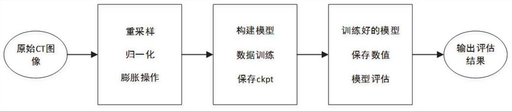
[0055] The processing module is used to resample, normalize and carry out expansion operation processing to the obtained lung CT scan image to be detected;

[0056] The detection module is used to process the processed lung CT scan image to be detected by using the trained detection model to obtain a detection result; the detection result includes whether there is a pulmonary nodule in the lung CT scan image to be detected, And the calibration position and area size of pulmonary nodules;

[0057] The trained detection model is obtained by using the training set; the training set includes a plurality of lung CT scan images, and labels of lung nodule positions and regions in the marked images.

[0058] In the present embodiment 1, utilize above-mentioned pulmonary nodule det...

Embodiment 2

[0081] In the present embodiment 2, provide a kind of pulmonary nodule detection system, this system comprises:

[0082] An acquisition module, configured to acquire a lung CT scan image to be detected;

[0083] The processing module is used to resample, normalize and carry out expansion operation processing to the obtained lung CT scan image to be detected;

[0084] The detection module is used to process the processed lung CT scan image to be detected by using the trained detection model to obtain a detection result; the detection result includes whether there is a pulmonary nodule in the lung CT scan image to be detected, And the calibration position and area size of pulmonary nodules;

[0085] The trained detection model is obtained by using the training set; the training set includes a plurality of lung CT scan images, and labels of lung nodule positions and regions in the marked images.

[0086] Such as figure 1 As shown, in the present embodiment 2, utilize above-men...

Embodiment 3

[0130] Embodiment 3 of the present invention provides a non-transitory computer-readable storage medium, the non-transitory computer-readable storage medium is used to store computer instructions, and when the computer instructions are executed by a processor, the pulmonary nodule detection method is implemented instruction, the method includes:

[0131] Obtain a CT scan image of the lung to be detected;

[0132] Resampling, normalizing and expanding the mask for the acquired lung CT scan image to be detected;

[0133] Use the trained detection model to process the processed lung CT scan image to obtain the detection result; the detection result includes whether there are pulmonary nodules in the lung CT scan image to be detected, and the number of pulmonary nodules Calibration position and area size;

[0134] The trained detection model is obtained by using the training set; the training set includes a plurality of lung CT scan images, and labels of lung nodule positions an...


PUM


Abstract

The invention provides a pulmonary nodule detection method and system, and belongs to the technical field of image recognition, and the method comprises the steps of obtaining a to-be-detected lung CT scanning image; performing resampling and normalization on the obtained lung CT scanning image to be detected, and performing expansion operation processing on the mask; processing the processed to-be-detected lung CT scanning image by using the trained detection model to obtain a detection result, wherein the detection result comprises whether the pulmonary nodule exists in the to-be-detected lung CT scanning image and the calibration position and the area size of the pulmonary nodule, the trained detection model is obtained through training by using a training set, the training set comprises a plurality of lung CT scanning images and labels for labeling positions and areas of pulmonary nodules in the images. According to the invention, accurate detection of the pulmonary nodule area is realized, the detection efficiency is improved, the detection quality is ensured, and the stability and efficiency of assisting doctors in disease diagnosis are improved.

Description

technical field [0001] The invention relates to the technical field of image recognition, in particular to a method and system for detecting pulmonary nodules. Background technique [0002] Early detection and treatment of potential small lesions of lung cancer play an important role in reducing the morbidity and mortality of lung cancer patients. At present, low-dose computed tomography (LDCT) is mainly used for lung nodule screening, and enhanced CT images are the direct basis for doctors to diagnose lung cancer through direct observation. The type, structure, size, and location of pulmonary nodules in CT images are different. It is difficult for doctors to process and analyze image data in screening slices, and they are more easily affected by the subjectivity of doctors. At the same time, the number of enhanced CT images is also increasing. work poses great challenges. [0003] Obviously, it is extremely difficult to observe and find tiny pulmonary nodules in slices on...

Claims


Application Information

Patent Timeline
Patent Type & Authority Applications(China)
IPC IPC(8): G06T7/00G06K9/62
CPCG06T7/0012G06T2207/10081G06T2207/30064G06F18/214
Inventor 万洪林赵莹莹王嘉鑫王晓敏
Owner SHANDONG NORMAL UNIV
Why Eureka
Try Eureka