Application of ASB17 in preparation of medicine for treating TRAF6 related inflammatory diseases

A technology for inflammatory diseases and drugs, applied in the field of ASB17 in the preparation of TRAF6-related inflammatory disease drugs, can solve problems such as no research

Pending Publication Date: 2021-12-10
佛山病原微生物研究院
3 Cites 0 Cited by

AI-Extracted Technical Summary

Problems solved by technology

[0005] Through the above analysis, the problems and defects of the existing technology are: the regulation mode of ASB17, and how to down-regulate the expres...

Abstract

The invention belongs to the technical field of biological medicine, and discloses application of ASB17 in preparation of a medicine for treating TRAF6 related inflammatory diseases, the Human ASB17 Sequence. Txt sequence of the ASB17 is shown as SEQ ID NO: 1, and the Mouse ASB17 Sequence. Txt sequence of the ASB17 is shown as SEQ ID NO: 2; the method for verifying ASB17 to prepare the medicine for treating the TRAF6 related inflammatory diseases comprises the following steps: separating mouse primary cells BMDCs and BMDMs; carrying out western blotting, co-immunoprecipitation and immunofluorescence; and protein stability detection and ubiquitination experiment. Through research and speculation, ASB17 and TRAF6 interact with each other, so that ubiquitination of TRAF6K48 can be inhibited, TRAF6 can be stabilized, and TRAF6-mediated inflammatory response can be promoted.

Application Domain

AntipyreticAnalgesics +3

Technology Topic

UbiquitinImmunoprecipitation +10

Image

  • Application of ASB17 in preparation of medicine for treating TRAF6 related inflammatory diseases
  • Application of ASB17 in preparation of medicine for treating TRAF6 related inflammatory diseases
  • Application of ASB17 in preparation of medicine for treating TRAF6 related inflammatory diseases

Examples

  • Experimental program(1)

Example Embodiment

[0088] In order to make the object, technical solution and advantages of the present invention more clear, the present invention will be further described in detail below in conjunction with the examples. It should be understood that the specific embodiments described here are only used to explain the present invention, not to limit the present invention.
[0089] In view of the problems existing in the prior art, the present invention provides a use of ASB17 in the preparation of TRAF6-related inflammatory disease medicines. The present invention will be described in detail below with reference to the accompanying drawings.
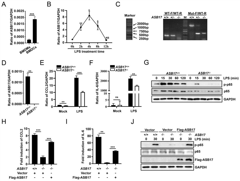
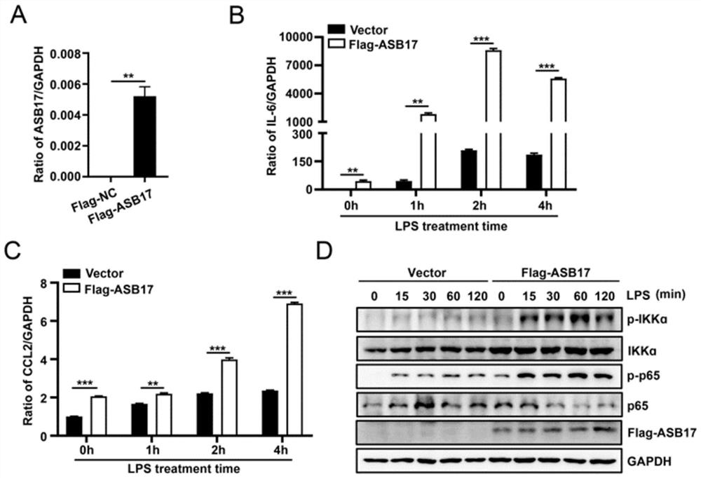
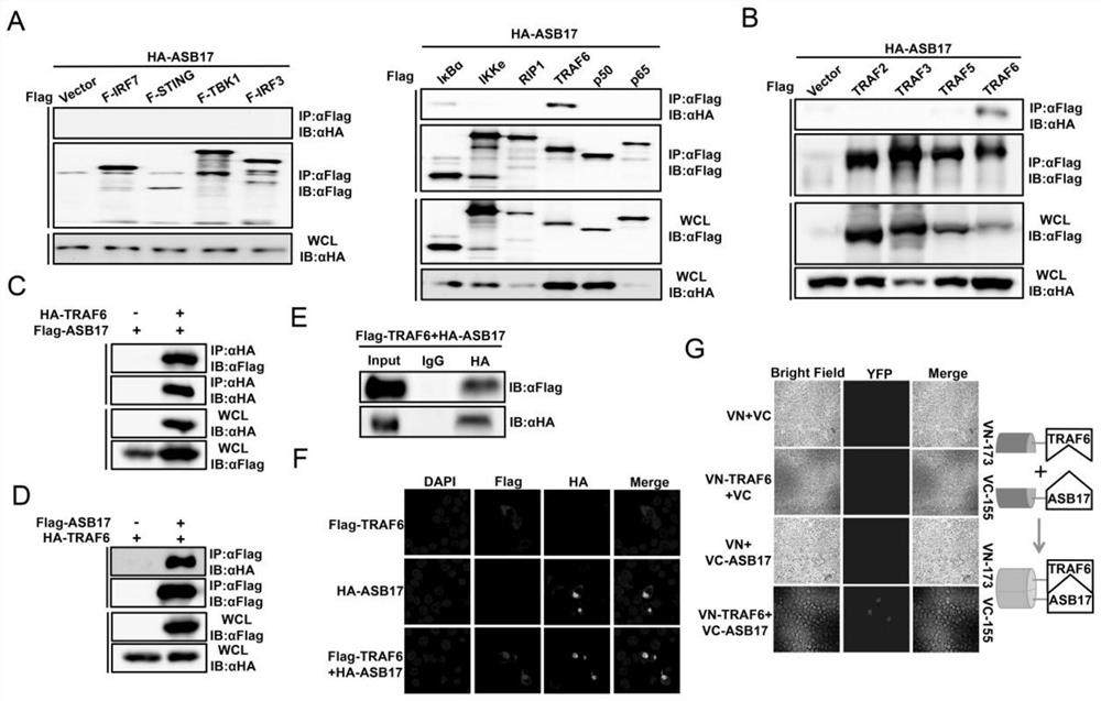
[0090] like Image 6 As shown, the method for verifying that ASB17 prepares a drug for treating TRAF6-related inflammatory diseases provided by the embodiments of the present invention includes the following steps:
[0091] S101, the isolation of primary mouse cells BMDCs and BMDMs;
[0092] S102, Western blotting, co-immunoprecipitation, and immunofluorescence;
[0093] S103, protein stability detection and ubiquitination experiment.
[0094] The technical solutions of the present invention will be further described below in conjunction with specific embodiments.
[0095] 1, the present invention analysis work:
[0096] (1) ASB17 can interact with TRAF6;
[0097] (2) At the cellular level, ASB17 was found to inhibit TRAF6 ubiquitination and stabilize TRAF6, while promoting TRAF6-mediated inflammatory response. On the basis of the experimental results and research progress at home and abroad, the specific mechanism and role of ASB17 in dendritic cells to promote TRAF6-mediated inflammatory response will be explored, which will provide an experimental basis for the host to promote TRAF6-mediated inflammatory response, and provide evidence for the role of TRAF6 in inflammatory response. Provide a theoretical basis for the regulation of TRAF6 and provide new ideas for the prevention and treatment of TRAF6-related diseases.
[0098] (1) Significance of the present invention:
[0099]Dendritic cells (DCs) are key sentinel cells and antigen-presenting cells (APCs) of the immune system. They link innate and adaptive immune responses and play an integral role in host defense against invading pathogens, including viruses, bacteria and parasites. Upon sensing pathogens via Toll-like receptors (TLRs), activation of DCs is primarily mediated by pattern recognition receptor/nuclear factor-κB (NF-κB) signaling and depends on proper ubiquitination of the corresponding signaling molecules. However, the ubiquitination and deubiquitination enzymes involved and their relationship to each other are not fully understood. TRAF6 is an important regulatory protein in the process of activating NF-κB transcriptional activity through Toll-like receptors, and its own polyubiquitination is quite important in this process. At present, several studies have shown that K48-linked TRAF6 ubiquitination can promote the degradation of TRAF6, thereby effectively blocking TLR-dependent inflammatory signaling pathways. Nonetheless, the molecular mechanisms regulating K48 ubiquitination and TRAF6 protein levels in TLRs-dependent innate immune responses remain unclear. How to regulate TRAF6-related inflammatory diseases has become a scientific research problem that researchers are committed to solving. This invention reveals the specific mechanism and function of ASB17 in dendritic cells to promote TRAF6-mediated inflammatory response, and provides experimental basis for the host to promote TRAF6-mediated inflammatory response , to provide a theoretical basis for enriching the regulatory mechanism of TRAF6-mediated inflammatory response, and to provide new ideas for the prevention and treatment of TRAF6-related diseases.
[0100] (2) object of the present invention
[0101] The research objective of the present invention is very clear, and two aspects of research are carried out closely around the specific mechanism and function of ASB17 promoting TRAF6-mediated inflammatory response.
[0102] The innovation of the present invention lies in that a new mechanism of ASB17 promoting TRAF6-mediated inflammatory response is proposed;
[0103] The present invention is a study on the molecular mechanism of host protein promoting TRAF6-mediated inflammatory response, and it is found that ASB17 can interact with TRAF6 through co-immunoprecipitation experiments. At the cellular level, elucidate the specific mechanism by which ASB17 promotes TRAF6-mediated inflammatory responses in dendritic cells. This provides a new mechanism for revealing host proteins to promote TRAF6-mediated inflammatory responses.
[0104] Novel proteins that regulate inflammation are also provided.
[0105] The invention clarifies the mechanism of ASB17 promoting TRAF6-mediated inflammatory response, and provides anti-inflammatory new protein for TRAF6 and related diseases.
[0106] 2. Content
[0107] 2.1 Effect of ASB17 promoting TRAF6-mediated inflammatory response in dendritic cells
[0108] By isolating primary mouse cells (BMDCs), it was found that ASB17 in dendritic cells can promote TRAF6-mediated inflammatory response through cell stimulation experiments.
[0109] 2.2 To clarify the specific mechanism of ASB17 promoting TRAF6-mediated inflammatory response
[0110] (1) ASB17 interacts with TRAF6 and can stabilize TRAF6
[0111] The interaction between ASB17 and TRAF6 was confirmed by co-immunoprecipitation and immunofluorescence; ASB17 was able to stabilize TRAF6 protein by cycloheximide chase analysis.
[0112] (2) ASB17 promotes TRAF6-mediated inflammatory response by stabilizing TRAF6 protein
[0113] Lentivirus overexpressed mASB17, and through lentivirus infection of BMDCs and Western blot experiments, it was confirmed that ASB17 promoted TRAF6-mediated inflammatory response by stabilizing TRAF6 protein.
[0114] 3. Experimental method
[0115] (1) Extraction of cell and tissue RNA and real-time fluorescent quantitative PCR
[0116] 1) The samples of the present invention are divided into cells and mouse tissues. When processing cell samples, if they are adherent cells, you can directly add 1ml of Trizol lysate to the cell culture dish, and repeatedly blow and beat with a pipette gun to make the cells fully understand. For suspension cells, first collect the cells by low-speed centrifugation, discard the supernatant and add 1ml Trizol to fully lyse the cells; when processing mouse tissue samples, first cut the fresh mouse tissue into pieces, and then take various tissues of appropriate size to Add 1ml of Trizol lysate to the RNase-free EP tube, and use a tissue grinder to fully break up the tissue cells.
[0117] 2) The above sample was fully lysed in Trizol lysate, and then placed at room temperature for 5 minutes to separate the protein-nucleic acid complex in the lysate.
[0118] 3) Add 200 μl of chloroform to the EP tube of 2, pay attention to cover the tube cap well, shake and mix well, and then place it at room temperature for 2 minutes.
[0119] 4) The above sample is centrifuged at 4°C at the highest speed for 10 minutes. After the centrifugation, the present invention will find that the sample in the EP tube is divided into three layers from bottom to top: red transparent layer, white precipitate layer and colorless transparent layer, and RNA is in the upper layer In a colorless transparent layer. Carefully transfer the colorless, clear liquid from the upper layer to a new RNase-free EP tube.
[0120] 5) Add 70% ethanol (prepared with DEPC water) equal to the volume of the above colorless transparent liquid into the EP tube of 4, and mix well by inverting up and down.
[0121] 6) Put the adsorption column in the kit into the collection tube, add the solution in 5 to the adsorption column, up to 700 μl each time, if it is not added at one time, it should be added until it is finished. Then the collection tube was kept at room temperature, centrifuged at 12000rpm for 30s, and the liquid in the collection tube was discarded.
[0122] 7) Add 700 μl of rinse solution 1 to the adsorption column, centrifuge at room temperature at 12,000 rpm for 30 s, and discard the liquid in the collection tube.
[0123] 8) Add 500 μl of rinse solution 2 (with absolute ethanol added) into the adsorption column, centrifuge at 12000 rpm for 30 s, discard the waste liquid in the collection tube, and then repeat the above operation once.
[0124] 9) After washing with the rinsing solution, centrifuge at the maximum speed for 2 minutes at room temperature, insert the adsorption column into a new RNase-free EP tube, and let stand at room temperature for several minutes to completely volatilize the ethanol on the adsorption column.
[0125] 10) After the ethanol is completely volatilized, add 30-50 μl DEPC water to the adsorption column, let it stand for 1 min, centrifuge at the maximum speed at room temperature for 1 min, and dissolve the RNA in the water at the bottom of the tube, measure the RNA concentration and record it.
[0126] 11) According to the reverse transcription system, add about 1 μg RNA at a time, 4 μl reverse transcriptase mixture 5×HiScript II qRTSuperMix, and make up to 20 μl with DEPC water. After mixing, centrifuge, put into the PCR instrument, set the program: 50°C for 15min, 85°C for 15s. Obtain the cDNA of the sample.
[0127] 12) According to the Q-PCR system, the present invention prepares the premix solution. Each well of the Q-PCR plate contains 1 μl of cDNA, 1 μl of forward and reverse primers, 10 μl of 2×ChamQ SYBR qPCR Master Mix and 8 μl of ddH 2 O. Make sure that each sample has three parallel control wells, which are tested on the machine after centrifugation.
[0128] (2) Cell culture and transfection
[0129] More common cell lines are used in the present invention. Dulbecco Modified Eagle Medium (DMEM) medium (which contains 10% fetal bovine serum FBS, 100 μg/ml streptomycin and 100 U/ml ampicillin) was used to culture HEK293T cells and Hela cells. HupG2 cells, Huh7 cells, U251 cells, A549 cells, primary BMDMs, primary BMDCs and RD cells were cultured in supplemented with 10% fetal bovine serum FBS, 1% double antibody (100μg/ml streptomycin and 100U/ml ampicillin penicillin) in DMEM medium. Roswell Park Memorial Institute (RPMI) 1640 medium (containing 10% fetal bovine serum FBS and 1% double antibody) was used to culture THP-1 cells. GC-1, GC-2, TM3 and TM4 cells were cultured in DMEM/F12 (GIBCO) medium supplemented with 10% FBS, 100 u/ml penicillin and 100 μg/ml streptomycin sulfate.
[0130] cell transfection
[0131] In the present invention, a large number of cell transfection experiments were carried out in HECK293T cells and Hela cells. Their transfection process is roughly the same, the steps are as follows:
[0132] 1) After determining the growth rate of the cells, spread the required cells into corresponding culture dishes (24-well plate, 12-well plate, 6-well plate, 600mm dish and 10cm dish) the night before.
[0133] 2) Make sure that the cell density is in the range of 60% to 80% before doing the experiment.
[0134] 3) Operate in an ultra-clean workbench, mix the prepared plasmid and transfection reagent with an equal amount of OPTI-MEM respectively, and the amount of OPTI-MEM added is 20% of the volume of the medium in the transfected culture dish. one-third. Wherein, according to the difference of the transfection reagent, the amount of the added plasmid and the transfection reagent are also different. Generally, when using Lipo 2000 for transfection, 1 μg of plasmid needs to be added with 2 μl of Lipo 2000. If PEI is used for transfection, 1 μg of plasmid needs to be added with 3 μl of PEI.
[0135] 4) Mix the plasmid mixture and the transfection reagent mixture evenly. If the transfection reagent is Lipo 2000, just let it stand at room temperature for 5 minutes; if transfect with PEI, let it stand at room temperature for 20 minutes.
[0136] 5) Use a pipette to evenly add the mixture in step 4 to the cell culture dish, and shake the culture dish properly to make it more uniform.
[0137] 6) After about 6 hours, replace the cells with fresh medium, and then incubate at 37°C, 5% CO 2 cultured in a cell culture incubator. It should be noted that the medium of HECK293T cells can be changed before transfection, but because Lipo 2000 is highly toxic to Hela cells, it must be replaced with fresh medium 4-6 hours after transfection.
[0138] (3) Isolation of mouse primary cells
[0139] Isolation of mouse bone marrow-derived dendritic cells (BMDCs)
[0140] 1) Wild mice and ASB17 knockout mice (6-8 weeks old) were prepared, all femurs and tibias were taken out under aseptic conditions, soaked in 70% alcohol for 5 minutes, and washed twice with PBS.
[0141] 2) Rinse the bone marrow with a syringe (drawing PBS) into a culture dish until the bone turns white; filter the suspension (200 mesh nylon mesh), centrifuge again, and resuspend with RPMI 1640 medium (containing 20ng/ml GM-CSF), Spread evenly onto 100mm cell culture dishes.
[0142]3) On day 3, add 10ml RPMI 1640 medium (containing 20ng/ml GM-CSF) to the culture dish; GM-CSF) to resuspend the cell pellet, and then put the cell suspension back into the original cell culture dish; BMDCs were obtained on the 10th day.
[0143] Isolation of mouse bone marrow-derived macrophages (BMDMs)
[0144] 1) Prepare wild mice and ASB17 knockout mice (6-8 weeks old), and remove all femurs and tibias under sterile conditions.
[0145] 2) Soak in 70% ethanol for 5 minutes, rinse twice with PBS.
[0146] 3) Rinse the bone marrow with a syringe (drawing PBS) into a culture dish until the bone turns white; filter the suspension (200 mesh nylon mesh), centrifuge, and remove red blood cells with red blood cell lysate.
[0147] 4) Add RPMI 1640 medium (containing 10-20% L929 cell culture medium) into the culture dish.
[0148] 5) On the 3rd and 5th day, the RPMI 1640 medium (containing 10-20% L929 cell culture medium) was used to replace the medium, and the BMDMs were obtained on the 6th day.
[0149] (4) Western blot
[0150] 1) Preparation of cell samples: After discarding the culture medium in the culture dish, rinse the cells with 2ml of pre-cooled PBS for 2-3 times; add a small amount of trypsin to digest the cells (preferably at 37°C), and then add 1ml containing Stop the trypsinization in the medium of serum, and resuspend the cells; blow and blow the pre-cooled PBS to resuspend the cells, centrifuge at room temperature and low speed, suck away the PBS, add 300 μl cell lysate (including protease inhibition); the cell suspension is passed through an ultrasonic cell disruptor Ultrasonic treatment, from turbidity to clarification; centrifuge the sample at 4°C for 10 minutes, transfer the supernatant to a new 1.5ml EP tube, add 80μl 5×SDS Loading Buffer, mix well and heat at 100°C for 10 minutes, the protein sample preparation is complete .
[0151] 2) Western blotting: After gel electrophoresis and transfer, remove the NC membrane and block with PBST (PBS+Tween) buffer containing 5% skim milk for 45 minutes; wash with PBST three times (5 minutes each time); add primary antibody (diluted in PBST buffer), incubated overnight at 4°C; recovered primary antibody, washed 3 times with PBST (5 min each); added secondary antibody (prepared with PBST containing 2% skimmed milk), incubated at room temperature for 45 min; washed 3 times with PBST, 10min each time; color development and exposure.
[0152] (5) Co-immunoprecipitation
[0153] 1) 36 to 48 hours after transfection, collect cell samples, rinse the cells with pre-cooled PBS for 1-2 times, then trypsinize, then stop trypsinization with serum-containing medium, collect in 1.5ml EP tube After the cells were centrifuged at low speed, the supernatant was removed, and the cells were rinsed twice with pre-cooled PBS, and finally the cell pellet was obtained.
[0154] 2) According to the amount of cells, add 500 to 1000 μl of RIPA lysate (the formula is 0.05M Tris hydrochloric acid, 0.001M ethylenediaminetetraacetic acid, 0.15M sodium chloride, 1% NP-40, 5% glycerol, pH value is 7.4) As well as protease inhibitors, cells were lysed on a rotary shaker for 30 min in a refrigerator at 4°C.
[0155] 3) After the lysis, the remaining cells were ultrasonically disrupted, and then centrifuged at 12000 rpm for 10 min at 4°C.
[0156] 4) After centrifugation, divide the supernatant into two parts, add 100μl to a new 1.5ml EP tube, then mix with 25μl of 5×SDS Loading Buffer, heat at 100°C for 10min, and store at -20°C after rapid centrifugation save. About 900 μl of the remaining supernatant was added, and about 20 μl of Protein A/G beads washed with RIPA lysate were added thereto, and adsorbed at 4° C. on a rotary shaker for 2 hours.
[0157] 5) After the adsorption is completed, centrifuge at 1000g, and transfer the supernatant to a new 1.5ml EP tube with a pipette gun.
[0158] 6) Add 1 μg of immunoprecipitation antibody to the solution of 5, and shake overnight at 4° C. (12-14 hours).
[0159] 7) The next day, about 40 μl of Protein A/G beads washed with RIPA lysate were added to the solution of 6, and adsorbed on a rotary shaker at 4° C. for 2 hours.
[0160] 8) After 2 hours, centrifuge the above-mentioned EP tube at 4°C at 1000 g for 1 min, discard the supernatant, and then add 1 ml of RIPA eluent (the formula is 0.05M Tris hydrochloric acid, 0.001M ethylenediaminetetraacetic acid, 0.3M sodium chloride, 1% NP-40, 5% glycerin, the pH value is 7.4), after mixing up and down gently up and down, centrifuge at 1000g at 4°C for 1min, carefully suck the supernatant with the washing liquid gun, and repeat the elution 6 times.
[0161] 9) For the last centrifugation, use a large pipette tip to cover a 10 μl small pipette tip, and try to suck up the supernatant.
[0162] 10) Add 60 μl of 2×SDS Loading Buffer to the EP tube above, then heat at 100°C for 10 minutes, and after brief centrifugation, put the protein immunoprecipitation sample in a -20°C refrigerator.
[0163] 11) Finally, the expression of the target protein in the immunoprecipitation and cell lysate was detected by western blot experiment.
[0164] (6) Immunofluorescence
[0165] In HECK293T cells, the distribution of target proteins in the cells and whether they co-localize were observed by transfection or co-transfection of plasmids alone. After transfection, the medium in the confocal dish was discarded, and the cells were rinsed twice with pre-cooled PBS. The mixture of methanol and acetone at a ratio of 1:1 was pre-cooled at 4°C in advance, added to the confocal dish, and then placed at 4°C for 20 minutes to fix and permeabilize the cells. After the permeabilized cells were fixed by rinsing with PBS (3 times, 5 min each), 3% BSA (prepared in PBS) was added and blocked at room temperature for 1 h. After blocking, rinse with PBS (3 times, 5 min each time), then add the fluorescent primary antibody prepared with 3% BSA to the confocal dish, and incubate at 4°C overnight (12-16 h). After incubation with the primary antibody, rinse with PBS buffer (3 times, 10 min each time), add fluorescent secondary antibody prepared with 10% BSA to a confocal dish, and store in tinfoil at room temperature for 45 min in the dark. After incubation with the secondary antibody, rinse with PBS three times, each time for 10 minutes, add the nuclear dye DAPI (prepared in methanol) to the confocal dish, protect from light, and place at 37°C for 5 minutes. Finally, the cells were rinsed with PBS buffer solution for 3 times (10 min each time), and the samples could be observed with a laser confocal microscope.
[0166] (7) Ubiquitination experiment
[0167] 1) Collect cell samples 36 to 48 hours after cell transfection, for details, please refer to the operation of collecting cells by immunoprecipitation.
[0168] 2) Collect the cells into a 1.5 ml EP tube, add 100 μl RIPA lysate (containing 1% SDS), pipette repeatedly, and then heat at 100° C. for 5 minutes.
[0169] 3) Add 900 μl of RIPA lysate to the solution of 2. At this time, the present invention obtains 0.1% SDS, and then place it on ice for 20 minutes.
[0170] 4) The above-mentioned cells were ultrasonically disrupted, then centrifuged at 12000 rpm for 10 min at 4°C, and the supernatant was collected into a new 1.5ml EP tube.
[0171] 5) Pipette 100 μl of the liquid in the above 1.5ml EP tube into a new 1.5ml EP tube, add 25 μl 5×SDSLoading Buffer and mix well, heat at 100°C for 10min, centrifuge and store at -20°C.
[0172] 6) Add 20 μl of Protein A/G beads washed with RIPA lysate to the remaining liquid, and pre-adsorb for 2 hours.
[0173] 7) Centrifuge the above EP tube at 4°C at 1000g, transfer the supernatant to a new 1.5ml EP tube and add the corresponding antibody for IP. overnight at 4°C on a rotary shaker.
[0174] 8) Add 40 μl of washed Protein A/G beads and continue to adsorb for 2 hours.
[0175] 9) After the adsorption, the RIPA eluent elutes the above-mentioned Protein A/G beads. 4°C, 1000g rotating speed, repeated 6 times.
[0176] 10) For the last time, aspirate the supernatant as much as possible, then add 60 μl 2×SDS Loading Buffer and mix evenly, heat at 100°C for 10 minutes, and centrifuge briefly at high speed to complete the protein sample preparation.
[0177] 4. Experimental results and discussion
[0178] The transcription factor nuclear factor kappa enhancer binding protein (NF-κB) controls many physiological activities including inflammation and innate immunity. After being stimulated by lipopolysaccharide (LPS), Toll-like receptors activate a series of enzymatic reactions in cells, and then stimulate the transcriptional activity of NF-κB, which shows the importance of Toll-like receptors for the activation of NF-κB. TRAF6 is an important regulatory protein in the process of activating NF-κB transcriptional activity through Toll-like receptors, and its own polyubiquitination is very important in this process. At present, several studies have shown that K48-linked TRAF6 ubiquitin can promote the degradation of TRAF6, thereby effectively blocking the TLR-dependent inflammatory signaling pathway. Nonetheless, the molecular mechanisms regulating K48 ubiquitination and TRAF6 protein levels in TLR-dependent innate immune responses remain unclear. Ankyrin repeat and SOCS box containing protein 17 (ASB17) is a member of the ASB family. The current function of ASB17 is not very clear, and there are basically no reports in the literature. It is only known that it can be expressed in large quantities in the testicular organs of mice. The present invention found that ASB17 deficiency can inhibit LPS-induced NF-κB transcription activation activity, while overexpression of ASB17 can promote this process. At the same time, the present invention confirms that ASB17 can interact with TRAF6, and that ASB17 can inhibit the polyubiquitination of TRAF6 and stabilize the protein level of TRAF6. Judging that it may be an important inflammatory pathway regulatory protein in the host, which plays an important role in protecting the host from excessive activation of its own inflammatory response. Therefore, the present invention deeply explores the function of ASB17 in regulating TRAF6-mediated inflammatory response.
[0179] 4.1 ASB17 promotes LPS-mediated NF-κB activation in BMDCs
[0180] Previous studies have shown that ASB17 is mainly expressed in the testis. However, expression of this gene can be detected in bone marrow-derived macrophages (BMDMs) and dendritic cells (BMDCs) (see figure 1A). In particular, its expression level in BMDCs can be induced by LPS, an important pyrogen from Gram-negative bacteria (see figure 1 B). It suggested that ASB17 may be involved in the pathological process of infectious inflammation. In order to study the role of ASB17 in dendritic cells, the present invention uses ASB17 purchased from the company +/- Mice produce ASB17 -/- mice, and isolate their BMDCs (see figure 1 C, D). We found that knockdown of ASB17 significantly attenuated the expression of LPS-induced pro-inflammatory cytokines CCL2 and IL-6 in BMDCs (see Figure 1E,F). The NF-κB signaling pathway plays a key role in the production of pro-inflammatory cytokines when TLR4 is activated by LPS. By detecting this pathway, the present invention found that when LPS stimulated BMDCs, the loss of ASB17 significantly reduced the phosphorylation of NF-κB component p65 (see figure 1 G), showing inhibition of NF-κB activation. Furthermore, when ASB17 in ASB17 -/- When overexpressed in BMDCs, the present invention detected the induction of pro-inflammatory cytokines and the phosphorylation of NF-κB p65 to verify its specificity. ASB17 -/- Overexpression of ASB17 in BMDCs restored LPS-induced expression of CCL2 and IL-6 and activation of NF-κB p65 (see figure 1 H, I, J). The results of the present invention indicate that ASB17 is very important for LPS-mediated NF-κB activation.
[0181] 4.2 ASB17 promotes LPS-mediated NF-κB activation in THP1
[0182] In order to further study the role of ASB17 in NF-κB signal transduction, the present invention constructed THP-1 cells stably expressing ASB17 protein (see figure 2 A). Overexpression of ASB17 significantly promoted the expression of IL-6 and CCL2 regardless of LPS stimulation (see figure 2 B, C). Consistent with knockout experiments, overexpression of ASB17 significantly promoted NF-κB activation (see figure 2 D). In conclusion, ASB17 can promote LPS-mediated NF-κB activation.
[0183] 4.3 ASB17 can interact with TRAF6
[0184] In order to study how ASB17 regulates the NF-κB signaling pathway, the present invention screened the key molecules involved in or related to the pathway by immunoprecipitation, including IRF7, STING, TBK1, IRF3, IκBα, IKKε, RIP1, TRAF6, P50 and P65 , to determine which molecules might interact with ASB17. Among these molecules, the present invention found that only TRAF6 interacts with ASB17 (see image 3 A). In order to ensure the specificity of the interaction between TRAF6 and ASB17, the present invention also constructed TRAF family related genes TRAF2, TRAF3 and TRAF5 for immunoprecipitation. It was shown that ASB17 can specifically precipitate TRAF6 (see image 3 B). Further co-immunoprecipitation (Co-IP) and reciprocal co-immunoprecipitation analyzes confirmed the interaction of ASB17 and TRAF6 (see image 3 C, D, E). Confocal microscopy showed that ASB17 co-localized with TRAF6 in cells (see image 3 F). The present invention also uses a bimolecular fluorescence complementation (BiFC) assay system to detect such protein-protein interactions. The present invention constructs VN-173-TRAF6 and VC-155-ASB17 plasmids and transfects them into cells. No fluorescence was observed in the control group, but significant fluorescence was observed in the experimental group transfected with both VN-173-TRAF6 and VC-155-ASB17. The results indicated that ASB17 could interact with TRAF6 (see image 3 F). Collectively, these results suggest that ASB17 is physiologically linked to TRAF6. Given that TRAF6 plays a key role in NF-κB signal transduction, the present invention believes that ASB17 regulates NF-κB signal pathway through the interaction with TRAF6.
[0185] 4.4 ASB17 inhibits TRAF6 polyubiquitination and stabilizes TRAF6 protein
[0186] To explore how ASB17 affects the function of TRAF6, the present invention investigated TRAF6 protein levels. HEK293T was quantitatively transfected with TRAF6, EGFP (as a control) and different doses of ASB17. Western blotting analysis showed that ASB17 could increase the protein level of TRAF6, but did not affect the expression of EGFP (see Figure 4 A). In order to study the stability of TRAF6 protein, the present invention uses cycloheximide (CHX), which prevents the synthesis of cellular protein, to carry out protein decay test. The results showed that overexpression of ASB17 significantly reduced the degradation rate of TRAF6 protein, indicating that ASB17 stabilized TRAF6 protein (see Figure 4 B).
[0187] The present inventors then examined whether polyubiquitination of TRAF6 is regulated by ASB17. In HEK293T cells, ASB17 can significantly inhibit the polyubiquitination of TRAF6 (see Figure 4 C). Furthermore, ASB17 inhibited polyubiquitination of TRAF6 in an ASB17 dose-dependent manner (see Figure 4 D). A number of E3 ubiquitin ligases have been reported to target TRAF6-linked ubiquitin chains (K48-linked or K63-linked). The present invention constructs ubiquitin mutation vectors K48O and K63O (except for the lysine residues at positions 48 and 63, all lysine residues are changed to arginine residues). Ubiquitin assays revealed that ASB17 inhibits K48- and K63-linked polyubiquitination of TRAF6 (see Figure 4 E). To further ensure that ASB17 inhibits TRAF6 K48-linked polyubiquitination, the present inventors found that ASB17 can inhibit TRAF6 K48-linked polyubiquitination in a dose-dependent manner (see Figure 4 F). In addition, overexpression of ASB17 also increased endogenous TRAF6 protein levels in THP-1 (see Figure 4 G). Overexpression of ASB17 significantly inhibited LPS-induced TRAF6 polyubiquitination and K48-linked polyubiquitination in THP-1 (see Figure 4 h). Consistent with this, ASB17 knockdown decreased endogenous TRAF6 protein levels and increased TRAF6 polyubiquitination in BMDCs (see Figure 4 I, J). In conclusion, ASB17 can inhibit the polyubiquitination of TRAF6, suggesting that ASB17 protects TRAF6 from degradation to promote NF-κB activation.
[0188] 4.5 The aa177-250 fragment of ASB17 is required to interact with the zinc finger domain of TRAF6 and inhibit the polyubiquitination of TRAF6
[0189] TRAF6 contains RF domain, ZnF domain and TRAF-C domain (see Figure 5 A). Co-IP analysis indicated that ASB17 could interact with the ZnF domain of TRAF6 (see Figure 5 B). It has been reported that ASB17 mainly contains ANK-box domain and SOCS-box domain (see Figure 5 C). Co-IP analysis revealed that the aa177-250 fragment of ASB17 between the ANK-box domain and the SOCS-box domain is required for the interaction with TRAF6 (see Figure 5 D). In order to further study the effect of ASB17 interaction on TRAF6 polyubiquitination, the present invention carried out ASB17 truncated ubiquitin detection assay. The results showed that deletion of the aa177-250 fragment abolished the activity of ASB17 on TRAF6 polyubiquitination inhibition. These data suggest that ASB17 interacts with TRAF6 through its aa177-250 fragment and inhibits TRAF6 polyubiquitination.
[0190] In the description of the present invention, unless otherwise stated, the meaning of "plurality" is two or more; the terms "upper", "lower", "left", "right", "inner", "outer" , "front end", "rear end", "head", "tail", etc. indicate the orientation or positional relationship based on the orientation or positional relationship shown in the drawings, and are only for the convenience of describing the present invention and simplifying the description, rather than Nothing indicating or implying that a referenced device or element must have a particular orientation, be constructed, and operate in a particular orientation should therefore not be construed as limiting the invention. In addition, the terms "first", "second", "third", etc. are used for descriptive purposes only and should not be construed as indicating or implying relative importance.
[0191] The above is only a specific embodiment of the present invention, but the scope of protection of the present invention is not limited thereto. Anyone familiar with the technical field within the technical scope disclosed in the present invention, whoever is within the spirit and principles of the present invention Any modifications, equivalent replacements and improvements made within shall fall within the protection scope of the present invention.

PUM

no PUM

Description & Claims & Application Information

We can also present the details of the Description, Claims and Application information to help users get a comprehensive understanding of the technical details of the patent, such as background art, summary of invention, brief description of drawings, description of embodiments, and other original content. On the other hand, users can also determine the specific scope of protection of the technology through the list of claims; as well as understand the changes in the life cycle of the technology with the presentation of the patent timeline. Login to view more.
Why Eureka
Try Eureka