
Cervical cancer MRI image segmentation device and method

An image segmentation, cervical cancer technology, applied in the field of electronic information, can solve the problems of poor cervical cancer image effect, etc., to achieve the effect of improving segmentation accuracy, fast reasoning, and reducing the amount of parameters

Pending Publication Date: 2021-12-28
FOURTH MILITARY MEDICAL UNIVERSITY +1
View PDF0 Cites 2 Cited by
  • Summary
  • Abstract
  • Description
  • Claims
  • Application Information

AI Technical Summary

Problems solved by technology

[0004] Embodiments of the present disclosure provide a device and method for segmenting cervical cancer MRI images, which can solve the problem of poor results when segmenting cervical cancer images

Method used


Image

  • Cervical cancer MRI image segmentation device and method
  • Cervical cancer MRI image segmentation device and method
  • Cervical cancer MRI image segmentation device and method

Examples


Embodiment 1

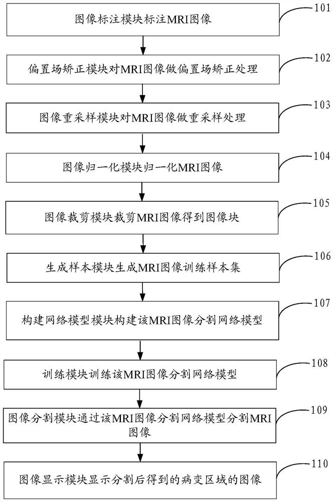
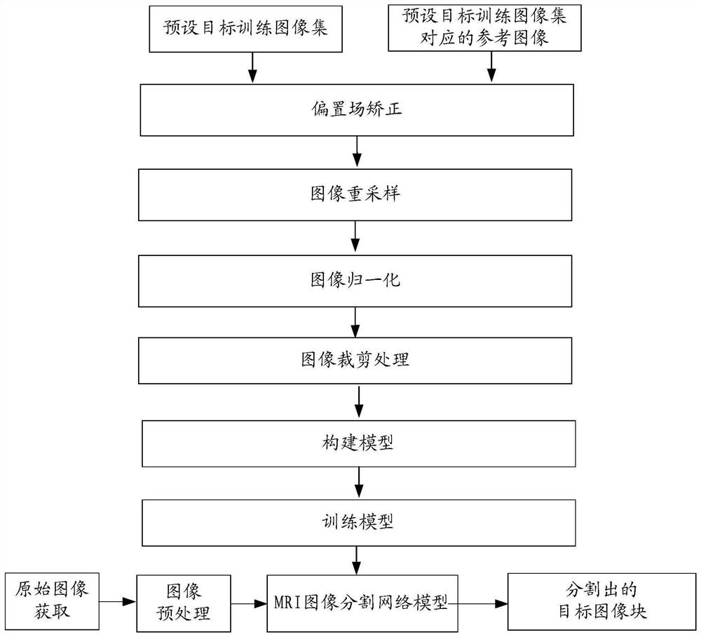
[0079] Embodiments of the present disclosure provide a method for segmenting cervical cancer MRI images, such as figure 1 and figure 2 As shown, the method is based on the MRI image segmentation network model of multi-view feature fusion, and the MRI image segmentation network model of multi-view feature fusion includes: a multi-view feature fusion module, which is used to extract features from different perspectives of the input image; The channel attention module is used to perform adaptive weighted fusion on the extracted features and accurately segment the MRI image according to the features after adaptive weighted fusion processing; specifically, the following steps are included:

[0080] 101. The image labeling module labels the MRI image.

[0081] The image annotation module in the method provided by the present disclosure annotates the MRI image, which specifically includes: the image annotation module outlines the lesion area frame by frame from the MRI image, and s...

Embodiment 2

[0148] An embodiment of the present disclosure provides a cervical cancer MRI image segmentation device, such as Figure 5As shown, the cervical cancer MRI image segmentation device 50 is based on a multi-view feature fusion MRI image segmentation network model, and the multi-view feature fusion MRI image segmentation network model includes: a multi-view feature fusion module, which is used to process input image blocks from different feature extraction from the angle of view; the channel attention module is used to perform adaptive weighted fusion on the extracted features and accurately segment the MRI image according to the features processed by the adaptive weighted fusion.

[0149] The device 50 includes an image labeling module 501, a bias field correction module 502, an image resampling module 503, an image normalization module 504, an image cropping module 505, an image segmentation module 506 and an image display module 507; wherein:

[0150] The image labeling module...


PUM


Abstract

The invention provides a cervical cancer MRI (Magnetic Resonance Imaging) image segmentation method and device, relates to the technical field of electronic information, and can solve the problem that the effect is poor when an image containing a cancer case is segmented. According to the specific technical scheme, when an MRI image including a cervical cancer lesion area is obtained, labeling processing, bias field correction processing and normalization processing are carried out on the MRI image, and the processed MRI image is input into an MRI image segmentation network model with multi-view feature fusion; according to the MRI image segmentation network model based on multi-view feature fusion, segmentation processing of an MRI image is realized through image interlayer feature extraction and image intra-layer feature extraction in the image, so that an image region containing a cervical cancer lesion region in the MRI image is obtained. The present disclosure is used for image segmentation processing.

Description

technical field [0001] The present disclosure relates to the field of electronic information technology, and in particular to a device and method for segmenting cervical cancer MRI images. Background technique [0002] Cervical cancer is one of the most common malignant tumors of the female reproductive system. The incidence rate is increasing, and the patients tend to be younger. MRI has a high image resolution for soft tissues, does not have ionizing radiation for patients, can perform imaging from different directions and sequences, and can clearly display the structure of the cervix, uterine body, vagina and surrounding tissues, and is often used in the clinical examination of cervical cancer . In actual clinical treatment, radiologists need to manually outline the area where cervical cancer is located in the MRI imaging system, and then formulate a detailed radiotherapy and chemotherapy plan based on the size and location of the outlined cervical cancer. Due to the la...

Claims


Application Information

Patent Timeline
Patent Type & Authority Applications(China)
IPC IPC(8): G06T7/11G06K9/46G06K9/62
CPCG06T7/11G06T2207/10088G06T2207/30096G06F18/253
Inventor 赵丽娜张晓鹏黄陆光张莹杨华刘波缑水平
Owner FOURTH MILITARY MEDICAL UNIVERSITY
Learn More