Supercharge Your Innovation With Domain-Expert AI Agents!

Protective device for imaging department

A protective device and image technology, which is applied in the field of medical equipment, can solve the problems of unusable protective equipment and easy damage of buckle-type protective equipment.

Inactive Publication Date: 2022-01-18
商丘市第一人民医院
View PDF0 Cites 1 Cited by
  • Summary
  • Abstract
  • Description
  • Claims
  • Application Information

AI Technical Summary

Problems solved by technology

[0005] In order to overcome the disadvantage that the buckle-type protective equipment is easy to be damaged when it is repeatedly worn and plugged in, thereby causing the entire protective equipment to be unusable, the present invention provides a protective device for the imaging department

Method used


Image

  • Protective device for imaging department
  • Protective device for imaging department
  • Protective device for imaging department

Examples


Embodiment 1

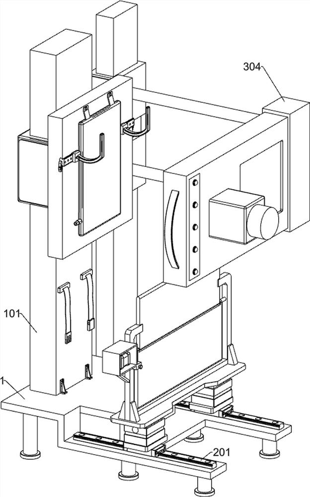
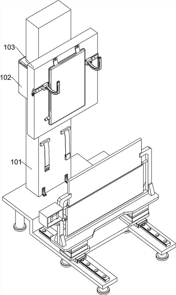
[0027] An imaging department protective device, such as Figure 1-6 As shown, it includes a bottom frame 1, an auxiliary support assembly, a protection assembly and a limit assembly; an auxiliary support assembly for supporting the human body is installed on the upper left side of the bottom frame 1; The protective component; the lower part of the right side of the auxiliary support component is equipped with a limit component for limit.

[0028] The auxiliary support assembly includes a first mounting plate 101, a first electric lifter 102, a fixed plate 103, an air bag 104, an adjustment slide rail 105, a translation slider 106, a first pin 107, an arc support handle 108, a ferrule 109 and The second bolt 110; the upper left part of the chassis 1 is fixedly connected with the first mounting plate 101; the first mounting plate 101 is equipped with the first electric lifter 102; the right side of the first electric lifter 102 is fixedly connected with the fixing plate 103 The...

Embodiment 2

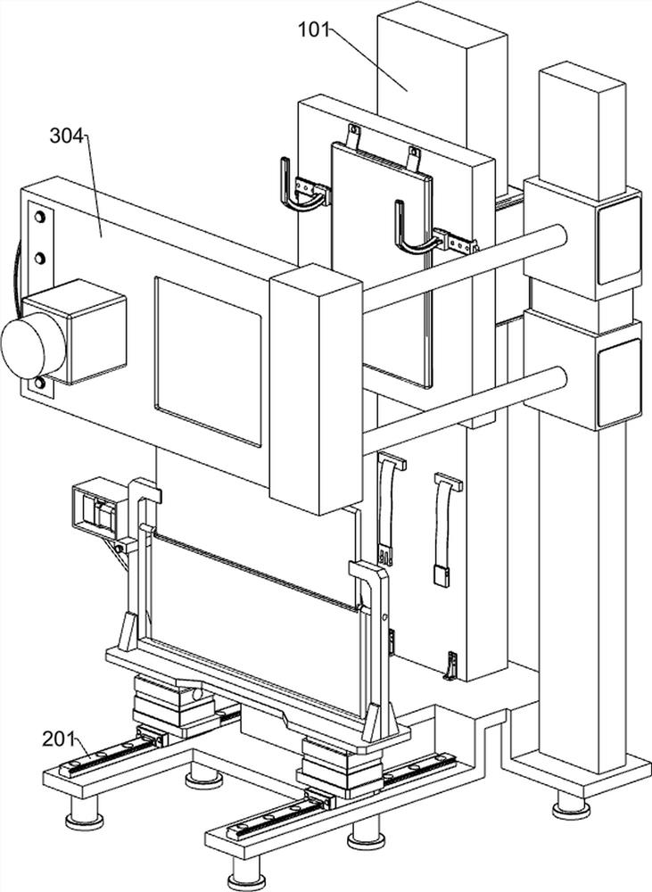
[0038] On the basis of Example 1, such as figure 1 and Figure 7 As shown, a chest X-ray assembly is also included, and the chest X-ray assembly is installed on the left side of the rear side of the chassis 1. The Chest X-ray assembly includes a second mounting plate 301, a second electric lifter 302, a fixed rod 303 and a chest X-ray instrument 304; A second mounting plate 301 is fixedly connected to the left side of the rear side of the chassis 1; two second electric lifters 302 are mounted on the second mounting plate 301; a fixed rod is fixedly connected to the right side of the two second electric lifters 302 303; the chest X-ray instrument 304 is fixedly connected to the right side of the two fixing rods 303.

[0039] After the examinee has adjusted his posture, the two second electric lifters 302 are controlled to lift up and down on the second mounting plate 301, and then the two fixed rods 303 are used to drive the chest X-ray instrument 304 up and down, so that the ...


PUM


Abstract

The invention relates to the field of medical instruments, in particular to a protective device for the imaging department. The invention solves the technical problem that a buckle type protective appliance is easy to damage when being repeatedly worn and inserted, so that the whole protective appliance cannot be used. According to the technical scheme, the protective device for the imaging department comprises a bottom frame, an auxiliary supporting assembly and the like; and the auxiliary supporting assembly used for supporting the human body is installed on the left portion of the upper side of the bottom frame. During use, protection is achieved through an adjustable protection plate separated from the human body, the protective appliance does not need to be contacted during use, the situation that the buckle type protective appliance is easy to damage when being repeatedly worn and inserted is avoided, auxiliary support is provided for people inconvenient in standing, people of different body types can be adapted, and the thoracic cavity of people can prop forwards and directly face an examination instrument in an examination process.

Description

technical field [0001] The invention relates to the field of medical equipment, in particular to an imaging department protective device. Background technique [0002] Chest X-ray is called fluoroscopy, which is a common X-ray examination method. X-ray examination is a double-edged sword. X-ray is harmful to human body, but many disease diagnoses cannot do without it. [0003] In the prior art, one-piece buckle-type protective equipment is usually used for protection during chest X-ray examinations. However, the hospital has a large number of people checking every day, and the buckle-type protective equipment is easily damaged when it is repeatedly worn and plugged in, which leads to the overall protection. The equipment cannot be used, resulting in a waste of resources. At the same time, the existing chest X-ray examination equipment has no auxiliary support for the examination of people who are inconvenient to stand, which makes it difficult to perform the examination. ...

Claims


Application Information

Patent Timeline
IPC IPC(8): A61B6/10A61B6/00
CPCA61B6/107A61B6/44A61B6/50
Inventor 隋愿张卿李玉舟
Owner 商丘市第一人民医院
Features
  • R&D
  • Intellectual Property
  • Life Sciences
  • Materials
  • Tech Scout
  • Unparalleled Data Quality
  • Higher Quality Content
  • 60% Fewer Hallucinations
Learn More