
Medical image diagnosis, comparison and reading device

A technology of film reading and imaging, applied in optical components, optics, instruments, etc., to achieve the effect of improving uniformity, smooth gloss, and easy installation or storage

Inactive Publication Date: 2022-03-18
永州职业技术学院
View PDF0 Cites 1 Cited by
  • Summary
  • Abstract
  • Description
  • Claims
  • Application Information

AI Technical Summary

Problems solved by technology

[0004] The purpose of the present invention is to provide a device for comparing and reading medical image diagnosis to solve the related problems raised in the above-mentioned background technology

Method used


Image

  • Medical image diagnosis, comparison and reading device
  • Medical image diagnosis, comparison and reading device
  • Medical image diagnosis, comparison and reading device

Examples


Embodiment 1

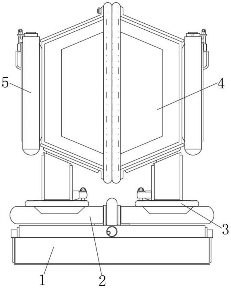
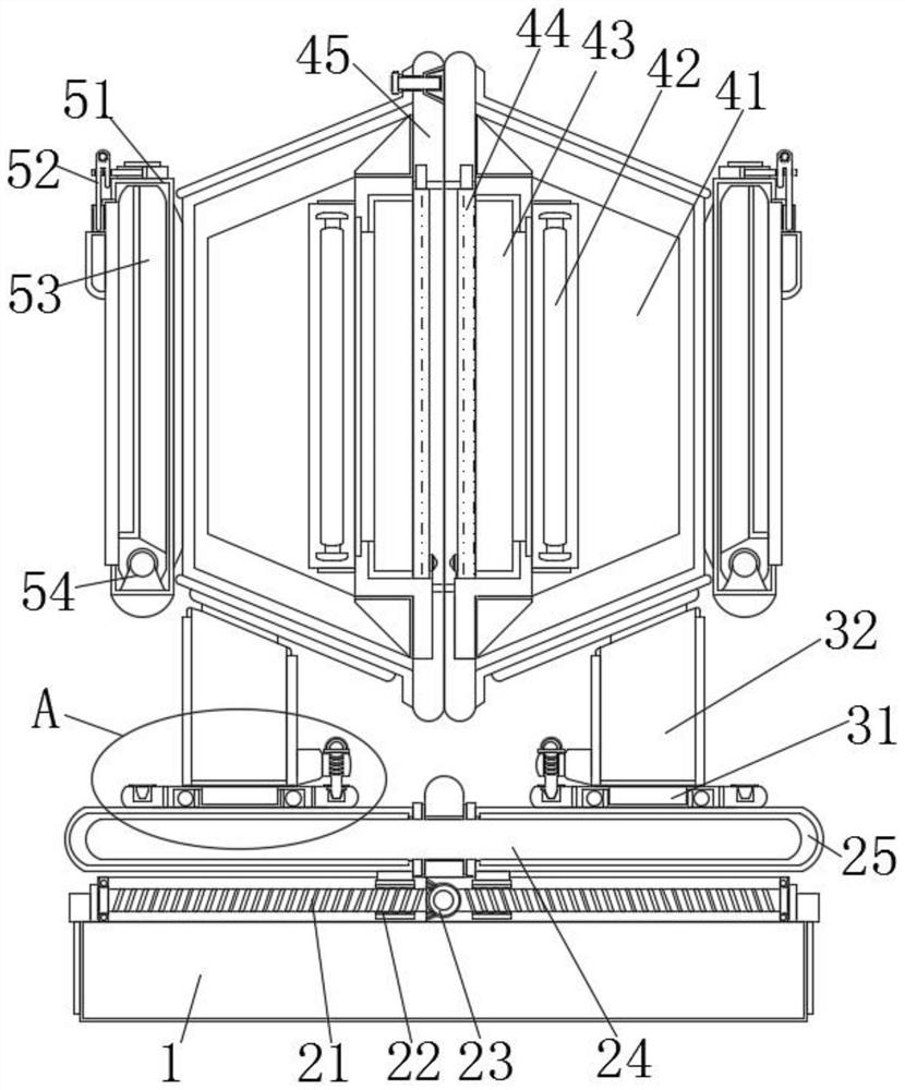
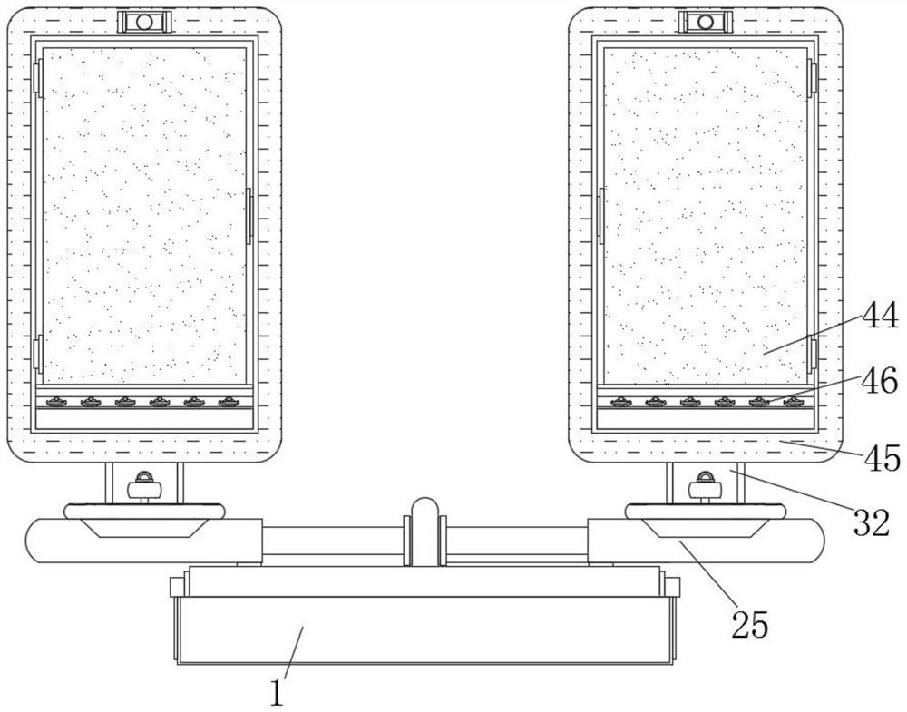
[0036] Example 1, such as Figure 1-3 As shown, when the installation groove 41 is unfolded and the film is observed, the sealing of the transparent acrylic plate 44 can be used to make the film a closed diagnostic comparison work, so as to avoid the dislocation of the film due to collision or the adhesion of external dust, and at the same time, when the medical personnel observe When there is an abnormality, the surface of the transparent acrylic plate 44 corresponding to the abnormality can be sucked and fixed through the transparent suction cup 46, so as to mark and record the abnormal points of the film, so as to facilitate subsequent judgment of the patient’s condition based on the marked records. targeted therapeutic work.

Embodiment 2

[0037] Example 2, such as figure 2 and Figure 7 As shown, before and after the film diagnosis and comparison, the film can be placed in the storage tank 53 in advance, so that the film is protected and stored under the seal of the storage tank 53 and the fixing tank 51, and the storage tank 53 can be positioned by the positioning member 52 , so that the film can be accommodated, and the two sets of films that need to be compared can also be fixed in the film groove 43 in advance, and the relative sealing of the two sets of installation grooves 41 can be used to protect the film well, so that In the follow-up work, the film can be diagnosed and observed by directly adjusting the distance and angle to increase the protection of the device.

[0038] Working principle: When the device is in use, first unfold the device in the storage state, and manually rotate the handle to drive the bevel gear set 23 to force the two-way screw rod 21 to rotate, and then force the two sets of i...


PUM


Abstract

A medical image diagnosis comparison reading device disclosed by the present invention comprises a mounting base, horizontal displacement mechanisms are arranged at the tops of the two sides in the mounting base, each horizontal displacement mechanism comprises a bidirectional screw rod, an inner threaded pipe, a bevel gear set, an inner sleeve column and an outer sleeve, the bidirectional screw rods are arranged at the tops of the two sides in the mounting base in a sleeving manner, and the bevel gear sets are arranged in the inner threaded pipes. A bevel gear set is arranged in the middle of the outer side of the two-way lead screw, inner threaded pipes matched with threads are symmetrically arranged on the outer side of the two-way lead screw, and the middle of the top of the mounting base is sleeved with an inner sleeve column. Through structural cooperation of the horizontal displacement mechanism and the angle adjusting mechanism, distance adjustment and angle adjustment of the two sets of mounting grooves can be provided, films arranged in the mounting grooves can better adapt to observation faces required by medical staff, and meanwhile the flexibility of film angle adjustment is improved through a 360-degree rotating system of the angle adjusting mechanism.

Description

technical field [0001] The invention relates to the technical field of medical equipment, in particular to a device for comparing and reading medical image diagnosis. Background technique [0002] The Department of Radiology has always been a traditional and necessary department in general hospitals, and has made great contributions to the hospital's medical and health research and teaching work. In the imaging diagnosis of the radiology department, X-ray films made by some X-optical instruments are often used to diagnose patients. The imaging observation device is generally a wall lamp. The film is inserted into the lower card slot of the wall lamp, and the wall lamp is turned on for observation. By observing the images on the imaging film to determine the patient's disease status, so as to carry out targeted treatment in the later stage. [0003] Most of the existing diagnostic devices place two sets of films in light boxes and adjust the angles of the two sets of light b...

Claims


Application Information

Patent Timeline
Patent Type & Authority Applications(China)
IPC IPC(8): G02B27/02G02B27/00
CPCG02B27/024G02B27/028G02B27/0006
Inventor 赵李湘蒋彬斌唐晓凤罗元元黄祥国
Owner 永州职业技术学院
Learn More