Method for simultaneously realizing three-dimensional subtraction arteriography, three-dimensional subtraction vein angiography and four-dimensional color angiography through image information post-processing of four-dimensional magnetic resonance angiography and medical imaging system

A technology of image information and magnetic resonance, applied in image data processing, detailed information related to graphical user interface, image analysis, etc., can solve problems such as difficult to observe, and achieve easy-to-evaluate effects

Pending Publication Date: 2022-04-08
韩国加图立大学校产学协力团 +1
View PDF0 Cites 0 Cited by
  • Summary
  • Abstract
  • Description
  • Claims
  • Application Information

AI Technical Summary

Problems solved by technology

However, there is a disadvantage that it is difficult to distinguish arteries and veins separately, so a solution for this is required

Method used


Image

  • Method for simultaneously realizing three-dimensional subtraction arteriography, three-dimensional subtraction vein angiography and four-dimensional color angiography through image information post-processing of four-dimensional magnetic resonance angiography and medical imaging system
  • Method for simultaneously realizing three-dimensional subtraction arteriography, three-dimensional subtraction vein angiography and four-dimensional color angiography through image information post-processing of four-dimensional magnetic resonance angiography and medical imaging system
  • Method for simultaneously realizing three-dimensional subtraction arteriography, three-dimensional subtraction vein angiography and four-dimensional color angiography through image information post-processing of four-dimensional magnetic resonance angiography and medical imaging system

Examples


Embodiment Construction

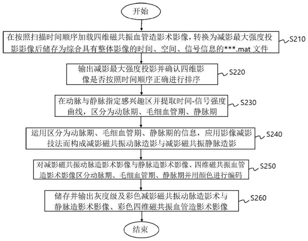
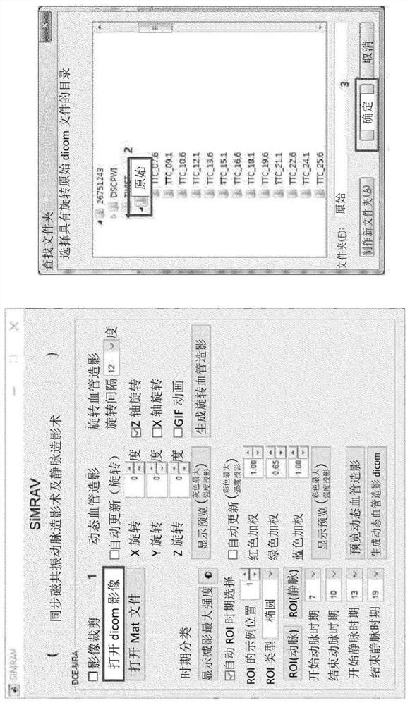
[0044] Hereinafter, preferred embodiments of the present invention will be described in detail with reference to the accompanying drawings. It should be understood that the embodiment described in this specification and the structure shown in the figure are only a preferred embodiment of the present invention, and not all represent the technical idea of ​​the present invention, so there may be alternatives from the perspective of this application Various equivalents and modifications thereof.

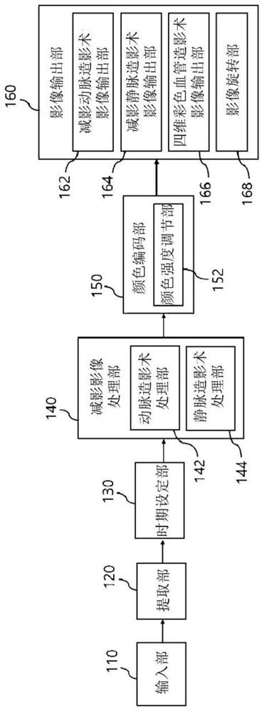
[0045] figure 1 It is a block diagram to represent the medical treatment for simultaneously realizing three-dimensional subtraction magnetic resonance angiography, three-dimensional subtraction magnetic resonance venography and four-dimensional color-coded magnetic resonance angiography through post-processing of image information of four-dimensional magnetic resonance angiography according to the present invention. An overall embodiment of an image system, including an input unit (110...


PUM


Abstract

The invention relates to a method and a medical image system for simultaneously realizing three-dimensional subtraction arteriography, three-dimensional subtraction vein angiography and four-dimensional color angiography through image information post-processing of four-dimensional magnetic resonance angiography.

Description

technical field [0001] The present invention relates to a post-processing of magnetic resonance angiography (MR Angiography) images, in particular to realizing three-dimensional subtraction angiography (3D Subtraction Arteriography) simultaneously through post-processing of image information of four-dimensional magnetic resonance angiography (4D MR Angiography). MR Arteriography), three-dimensional subtraction venography (3D SubtractionMR Venography) and four-dimensional color angiography (four-dimensional color-coded magnetic resonance angiography (Color-coded4D MR Angiography)) methods and medical imaging systems. Background technique [0002] As medical imaging devices mainly used for cerebrovascular diseases, there are Magnetic Resonance Imaging (MRI), Computed Tomography (CT), Single Photon Emission Computed Tomography (Single Photon Emission Computed Tomography, SPECT)), positron emission tomography (Positron Emission Tomography, PET) and ultrasound (Ultrasound), etc. ...

Claims


Application Information

Patent Timeline
Patent Type & Authority Applications(China)
IPC IPC(8): A61B5/00A61B5/055
CPCA61B5/055A61B5/0033A61B5/742G06T7/0012G06T2200/24G06T2207/10088G06T2207/10016G06T2207/20092G06T2207/30101G06T7/0016G06T19/20G06T2200/04G06T2207/10076G06T2207/20132G06T2207/20224G06T2207/30096G06T2219/2012
Inventor 金炫贞盧弘基
Owner 韩国加图立大学校产学协力团
Why Eureka
Try Eureka