Uterine myoma target image acquisition method based on residual network structure

A technology for uterine fibroids and target images, which is applied in the field of uterine fibroids target image acquisition based on a residual network structure, can solve the problems that the detection accuracy and speed of ultrasonic images cannot be taken into account at the same time, achieves generalization and strong applicability, and improves the Accuracy and simplicity

Pending Publication Date: 2022-04-26
SHUNDE HOSPITAL SOUTHERN MEDICAL UNIV (THE FIRST PEOPLES HOSPITAL OF SHUNDE FOSHAN) +1
View PDF0 Cites 1 Cited by
  • Summary
  • Abstract
  • Description
  • Claims
  • Application Information

AI Technical Summary

Problems solved by technology

[0005] Aiming at the problem that the accuracy and speed of ultrasonic image detection in the prior art cannot be taken into account at the same time, the present invention provides a method for acquiring a target image of uterine fibroids based on a residual network structure,

Method used


Image

  • Uterine myoma target image acquisition method based on residual network structure
  • Uterine myoma target image acquisition method based on residual network structure
  • Uterine myoma target image acquisition method based on residual network structure

Examples


Embodiment Construction

[0050] The present invention will be further described in detail below in conjunction with the embodiments and the accompanying drawings. It should be understood that the specific embodiments described here are only used to explain the present invention, not to limit the present invention. Based on the embodiments of the present invention, all other embodiments obtained by persons of ordinary skill in the art without making creative efforts belong to the protection scope of the present invention.

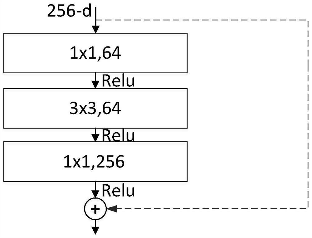
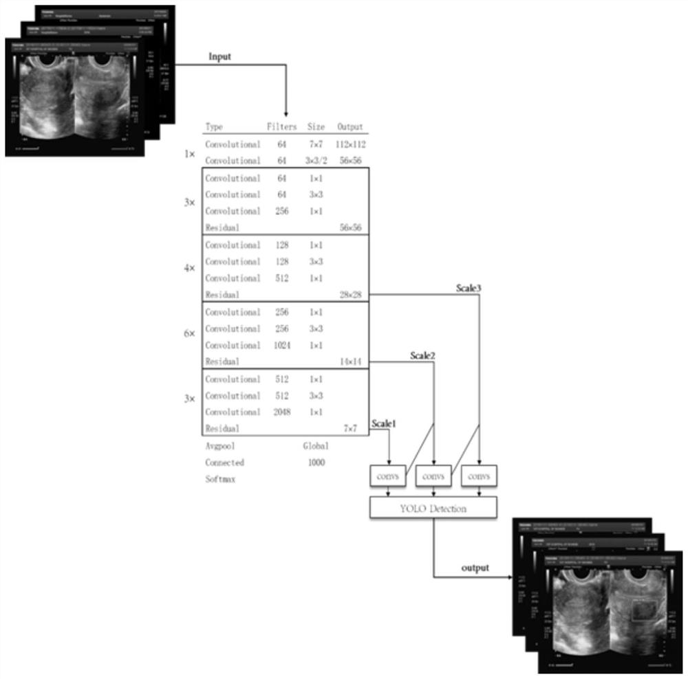
[0051] A uterine fibroid target image acquisition method based on residual network structure, the method includes the following two stages:

[0052] Phase 1, model training

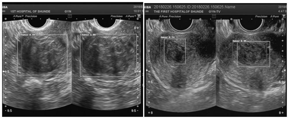
[0053] S1. On the ultrasound image of the original sample, mark the lesion target image area in the form of a rectangular frame on the area containing the uterine fibroid image to obtain a standard marking result, which includes a standard marking image and a standard marking file;

[0054]S2. The standard la...


PUM


Abstract

The invention is suitable for the technical field of medical image processing, and particularly relates to a hysteromyoma target image acquisition method based on a residual network structure, which comprises two stages of model training and model application. The model training comprises the following steps: S1, on an original sample ultrasonic image, carrying out lesion target image region labeling on a region containing a hysteromyoma image in a rectangular frame form; s2, the standard annotation image is detected through an improved YOLOv3 target detection model; and S3, results are unified, and a trained improved YOLOv3 target detection model is obtained. During application, a to-be-detected ultrasonic image is input into the trained improved YOLOv3 target detection model for detection, and a hysteromyoma target image area result is obtained. According to the method, the acquisition precision of the hysteromyoma image area can be improved, the detection speed is high, and the adaptability to small-target and multi-target tasks is high.

Description

technical field [0001] The invention relates to the technical field of artificial intelligence, in particular to a method for acquiring target images of uterine fibroids based on a residual network structure. Background technique [0002] Uterine fibroids often appear round or oval in ultrasound image data, with clear borders. Ultrasound manifestations of uterine fibroids are diverse. The reasons for the diversity include the following three aspects: first, the size and shape of uterine fibroids are complex; on the other hand, Due to the special imaging method of ultrasound imaging, the target of uterine fibroids in the image is similar to the gray scale of the background, and it is difficult to accurately delineate the boundary of the target area; Moreover, the contrast and hue of the image itself may vary due to differences in image acquisition equipment and environments. The diversity of uterine ultrasound images and targets makes it difficult to extract target features ...

Claims


Application Information

Patent Timeline
Patent Type & Authority Applications(China)
IPC IPC(8): G06T7/00G06N3/04G06N3/08
CPCG06T7/0012G06N3/08G06T2207/10132G06T2207/20081G06T2207/20084G06T2207/30096G06T2207/20132G06N3/045
Inventor 霍彤彤邓凯贤李丽欣叶哲伟吴蔚王子毅
Owner SHUNDE HOSPITAL SOUTHERN MEDICAL UNIV (THE FIRST PEOPLES HOSPITAL OF SHUNDE FOSHAN)
Why Eureka
Try Eureka