
Organ volume determination method and device, equipment and storage medium

A volume determination and organ technology, applied in the field of medical image processing, can solve problems such as organ errors and achieve the effect of improving accuracy

Pending Publication Date: 2022-04-26
CHISON MEDICAL TECH CO LTD
View PDF0 Cites 0 Cited by
  • Summary
  • Abstract
  • Description
  • Claims
  • Application Information

AI Technical Summary

Problems solved by technology

[0004] In the above scheme, the judgment of the organ volume depends on the influence of personal experience, and the size of the collected ultrasound image is related to the pose of the collection probe, and there is a large error in judging the volume of the organ manually

Method used


Image

  • Organ volume determination method and device, equipment and storage medium
  • Organ volume determination method and device, equipment and storage medium
  • Organ volume determination method and device, equipment and storage medium

Examples


Embodiment Construction

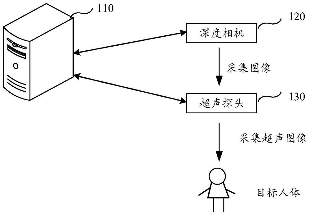
[0046] The technical solutions of the present application will be clearly and completely described below in conjunction with the accompanying drawings. Apparently, the described embodiments are some of the embodiments of the present application, not all of them. Based on the embodiments in this application, all other embodiments obtained by persons of ordinary skill in the art without making creative efforts belong to the scope of protection of this application.

[0047] It should be understood that the "indication" mentioned in the embodiments of the present application may be a direct indication, may also be an indirect indication, and may also mean that there is an association relationship. For example, A indicates B, which can mean that A directly indicates B, for example, B can be obtained through A; it can also indicate that A indirectly indicates B, for example, A indicates C, and B can be obtained through C; it can also indicate that there is an association between A an...


PUM


Abstract

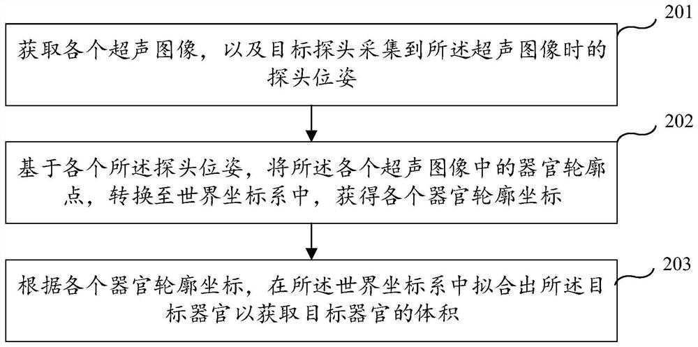
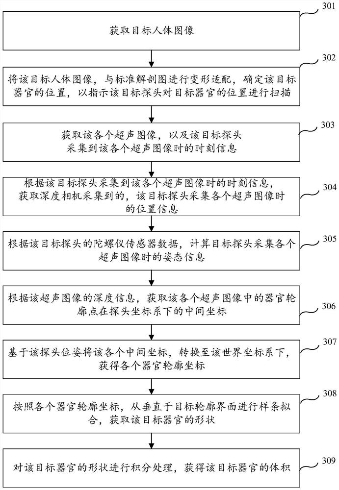
The invention relates to an organ volume determination method and device, equipment and a storage medium, and particularly relates to the technical field of computer vision. The method comprises the following steps: acquiring each ultrasonic image and a probe pose when a target probe acquires the ultrasonic image; on the basis of the probe poses, organ contour points in the ultrasonic images are converted into a world coordinate system, and contour coordinates of the organs are obtained; and fitting the target organ in the world coordinate system according to the contour coordinates of the organs to obtain the volume of the target organ. According to the scheme, the target organ is subjected to ultrasonic detection through different probe poses, so that organ contours in different directions can be calculated, the target organ can be well fitted to obtain the volume, the volume is not influenced by personal experience, and the accuracy of detecting the volume of the organ is improved.

Description

technical field [0001] The present invention relates to the field of medical image processing, in particular to a method, device, equipment and storage medium for determining an organ volume. Background technique [0002] Ultrasonic images reflect the differences in acoustic parameters in the medium, and can obtain information different from optics, X-rays, and y-rays. Ultrasound has a good ability to distinguish soft tissues of the human body, and can obtain useful signals with a dynamic range of up to 120dB, which is conducive to identifying small lesions in biological tissues. Ultrasound images can obtain the desired images without dyeing when displaying living tissues. [0003] In the actual medical field, when it is necessary to detect the organ volume of the human body, the doctor usually collects and processes the ultrasonic image of the human body to obtain the ultrasonic image of the human body. By comparison, it is judged whether there is any abnormality in the s...

Claims


Application Information

Patent Timeline
Patent Type & Authority Applications(China)
IPC IPC(8): G06T7/00G06T7/11G06T7/62G06T7/70G16H30/20
CPCG06T7/0012G06T7/70G06T7/62G06T7/11G16H30/20G06T2207/10136
Inventor 不公告发明人
Owner CHISON MEDICAL TECH CO LTD
Learn More