Method and device for screening diastolic and systolic images based on cardiac ultrasound video

A diastolic and systolic technology, applied in the field of screening diastolic and systolic images based on cardiac ultrasound video, can solve problems such as the inability to screen end-systolic and end-diastolic image quality and stability, and the impact of ejection fraction calculation accuracy, etc. To achieve the effect of stability assurance

Pending Publication Date: 2022-04-29
LEPU MEDICAL TECH (BEIJING) CO LTD
View PDF0 Cites 3 Cited by
  • Summary
  • Abstract
  • Description
  • Claims
  • Application Information

AI Technical Summary

Problems solved by technology

This method of operation relies too much on personal manual experience and proficiency in manual operation of equipment, and cannot effectively guarantee the quality and stability of screening images at the end of systole and end of diastole
The selected images will be used as the calculation basis for further calculation of ejection fraction, and in the case of poor image screening quality, it may also affect the calculation accuracy of ejection fraction

Method used


Image

  • Method and device for screening diastolic and systolic images based on cardiac ultrasound video
  • Method and device for screening diastolic and systolic images based on cardiac ultrasound video
  • Method and device for screening diastolic and systolic images based on cardiac ultrasound video

Examples


Embodiment Construction

[0048] In order to make the object, technical solution and advantages of the present invention clearer, the present invention will be further described in detail below in conjunction with the accompanying drawings. Obviously, the described embodiments are only some embodiments of the present invention, rather than all embodiments . Based on the embodiments of the present invention, all other embodiments obtained by persons of ordinary skill in the art without making creative efforts belong to the protection scope of the present invention.

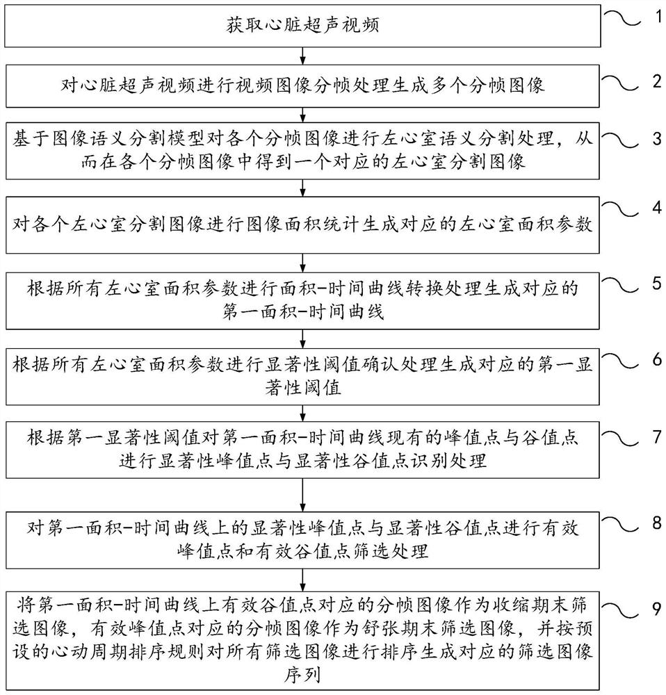
[0049] Embodiment 1 of the present invention provides a method for screening diastolic and systolic images based on cardiac ultrasound video, such as figure 1 As shown in the schematic diagram of a method for screening diastolic and systolic images based on cardiac ultrasound video provided in Embodiment 1 of the present invention, the method mainly includes the following steps:

[0050] Step 1, obtain cardiac ultrasound video.

[0051] H...


PUM


Abstract

The embodiment of the invention relates to a method and device for screening diastolic and systolic images based on a cardiac ultrasound video. The method comprises the following steps: acquiring the cardiac ultrasound video; performing video image framing to generate a plurality of framed images; performing left ventricle semantic segmentation on each framed image to obtain a corresponding left ventricle segmented image; performing image area statistics to generate corresponding left ventricular area parameters; performing area-time curve conversion to generate a first area-time curve; performing significance threshold confirmation to generate a first significance threshold; carrying out saliency peak and valley value point identification on the existing peak and valley value points of the first area-time curve; performing effective peak and valley value point screening on the significant peak and valley value points; and taking the framed image corresponding to the effective valley point as a screening image at the end of a contraction period, taking the framed image corresponding to the effective peak point as a screening image at the end of a relaxation period, and sorting all the screening images to generate a screening image sequence. According to the invention, the image screening quality stability can be ensured.

Description

technical field [0001] The invention relates to the technical field of data processing, in particular to a method and device for screening diastolic and systolic images based on cardiac ultrasound video. Background technique [0002] Ejection fraction refers to stroke volume, that is, the percentage of blood ejected from one side of the ventricle in one heartbeat to the end-diastolic volume of the ventricle, and is one of the important indicators for judging the health of the heart. The calculation of ejection fraction is related to end-systole and end-diastole ventricular volume. The real-time output of conventional echocardiography equipment is a set of video data, that is, echocardiographic video. When the operator plays this group of echocardiographic video on the device, he often screens multiple frames of end-systolic and end-diastolic images through personal experience for analysis. A reference image for the score. This method of operation relies too much on persona...

Claims


Application Information

Patent Timeline
Patent Type & Authority Applications(China)
IPC IPC(8): G06V20/40G06V10/26G06V10/46G06V10/82G06N3/08G06T7/00G06T7/41G06T7/62
CPCG06N3/08G06T7/0012G06T7/41G06T7/62G06T2207/10016G06T2207/10132G06T2207/20084G06T2207/20081G06T2207/30048
Inventor 李喆王思翰张碧莹曹君
Owner LEPU MEDICAL TECH (BEIJING) CO LTD
Why Eureka
Try Eureka