Medical image viewing method and device, equipment and storage medium

An image viewing and medical image technology, which is applied in the computer field, can solve the problems of large DICOM image files, reduce user viewing experience, and long user waiting time, and achieve the effects of reducing waiting time, improving viewing experience, and increasing viewing speed

Pending Publication Date: 2022-05-13
联仁健康医疗大数据科技股份有限公司
View PDF4 Cites 0 Cited by
  • Summary
  • Abstract
  • Description
  • Claims
  • Application Information

AI Technical Summary

Problems solved by technology

However, since the DICOM image file itself contains a sequence of images in groups, the DICOM image file is relatively large, such as more than 100MB, so it takes a long time to download the DICOM image file, and the user waits longer when the network quality is poor, reducing the User viewing experience

Method used


Image

  • Medical image viewing method and device, equipment and storage medium
  • Medical image viewing method and device, equipment and storage medium
  • Medical image viewing method and device, equipment and storage medium

Examples


Embodiment 1

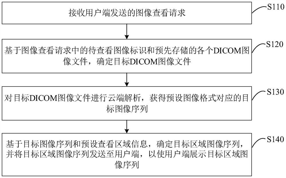
[0031] figure 1 It is a flow chart of a medical image viewing method provided by Embodiment 1 of the present invention. This embodiment is applicable to the situation of viewing two-dimensional medical images or three-dimensional medical images. The method can be executed by a medical image viewing device integrated in the cloud, and the device can be realized by software and / or hardware. Such as figure 1 As shown, the method specifically includes the following steps:

[0032] S110. Receive an image viewing request sent by the client.

[0033] Wherein, the user end may refer to a device end where a user views medical images. For example, the user end may refer to, but is not limited to, a web page end or an application end. Wherein, the webpage terminal may refer to a browser of a hospital doctor's workstation, and the application client may refer to a mobile phone client of a patient.

[0034] Wherein, the medical image may refer to a two-dimensional medical image or a t...

Embodiment 2

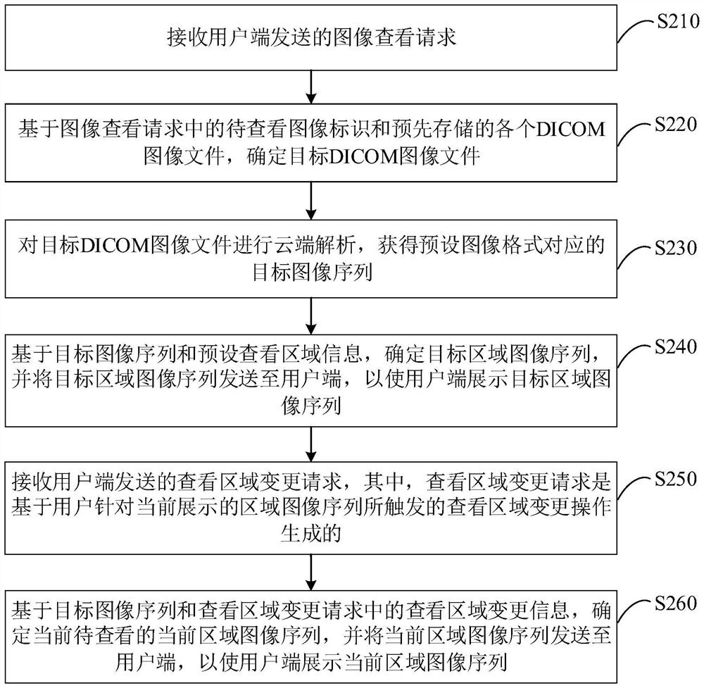
[0057] figure 2 It is a flowchart of a medical image viewing method provided by Embodiment 2 of the present invention. This embodiment describes in detail the process of changing the viewing area of ​​a medical image on the basis of the above embodiments. The explanations of terms that are the same as or corresponding to the above-mentioned embodiments will not be repeated here.

[0058] see figure 2 The medical image viewing method provided in this embodiment specifically includes the following steps:

[0059] S210. Receive an image viewing request sent by the client.

[0060] S220. Determine the target DICOM image file based on the identifier of the image to be viewed in the image viewing request and each pre-stored DICOM image file.

[0061] S230. Perform cloud analysis on the target DICOM image file to obtain a target image sequence corresponding to a preset image format.

[0062] S240. Based on the target image sequence and preset viewing area information, determine...

Embodiment 3

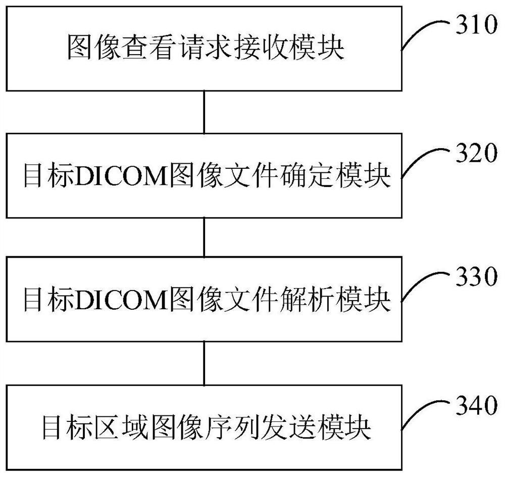
[0071] image 3 It is a schematic structural diagram of a medical image viewing device provided in Embodiment 3 of the present invention. This embodiment is applicable to viewing two-dimensional medical images or three-dimensional medical images. The device specifically includes: an image viewing request receiving module 310, Target DICOM image file determination module 320 , target DICOM image file parsing module 330 and target area image sequence sending module 340 .

[0072] Wherein, the image viewing request receiving module 310 is used to receive the image viewing request sent by the client; the target DICOM image file determination module 320 is used to determine based on the image identification to be viewed in the image viewing request and each DICOM image file stored in advance. Target DICOM image file; Target DICOM image file parsing module 330, for carrying out cloud analysis to target DICOM image file, obtains the corresponding target image sequence of preset image f...


PUM


Abstract

The embodiment of the invention discloses a medical image viewing method and device, equipment and a storage medium. The method comprises the following steps: receiving an image viewing request sent by a user side; determining a target DICOM image file based on a to-be-viewed image identifier in the image viewing request and each pre-stored DICOM image file; performing cloud analysis on the target DICOM image file to obtain a target image sequence corresponding to the preset image format; and based on the target image sequence and the preset viewing area information, determining a target area image sequence, and sending the target area image sequence to the user side, so that the user side displays the target area image sequence. Through the technical scheme of the embodiment of the invention, the viewing speed of the medical image can be improved, the waiting time of a user is shortened, and the viewing experience of the user is improved.

Description

technical field [0001] The embodiments of the present invention relate to computer technology, and in particular to a medical image viewing method, device, equipment and storage medium. Background technique [0002] With the rapid development of computer technology, doctors or patients can view medical images in electronic film form on terminal equipment, avoiding the disadvantages caused by paper films. [0003] At present, the medical images taken by patients are usually stored in the cloud in DICOM (Digital Imaging and Communications in Medicine) format. When a medical image of a patient needs to be viewed, the DICOM image file corresponding to the patient can be downloaded from the cloud to the client However, because the DICOM image file itself contains a sequence of images in groups, the DICOM image file is relatively large, such as more than 100MB, so it takes a long time to download the DICOM image file, and the user waits for a long time when the network quality is ...

Claims


Application Information

Patent Timeline
Patent Type & Authority Applications(China)
IPC IPC(8): G16H30/20
CPCG16H30/20
Inventor 赵子健
Owner 联仁健康医疗大数据科技股份有限公司
Why Eureka
Try Eureka