
Knee joint X-ray image quality control method and device

A quality control method and knee joint technology, applied in the field of medical imaging, can solve the problems of low efficiency of visual inspection, multiple data, limited time for doctors, etc., to solve the difficulty of calculating quality control parameters, reduce the amount of data calculation, and reduce The effect of memory footprint

Pending Publication Date: 2022-06-07
HANGZHOU SHENRUI BOLIAN TECH CO LTD +2
View PDF1 Cites 0 Cited by
  • Summary
  • Abstract
  • Description
  • Claims
  • Application Information

AI Technical Summary

Problems solved by technology

The determination of traditional image quality is done by senior radiologists with the naked eye. This quality control method usually has the following disadvantages: the number of doctors is small, and the number of senior doctors is even less; the time of doctors is limited. The efficiency is too low; it is difficult to completely unify the standards among different doctors
The application number is 202010431833.9, and the invention patent titled "Medical Image Quality Control Method and Computer Readable Storage Medium" proposes an image quality control method based on instance segmentation. This method has the following disadvantages: each bone block needs to be (Tibia, Fibula, Patella) Annotate instance-level segmentation labels, the amount of labeling is huge, and more data is required; the segmentation results need to be converted into corresponding linear measurement results, such as how to convert the segmentation results of tibia and femur For the corresponding angle relationship, this is a relatively complicated and error-prone process; for severely occluded bone blocks, the performance of model segmentation directly reflects the final quality, such as the overlapping part of the fibula head and tibia is often blurred and difficult to locate

Method used


Image

  • Knee joint X-ray image quality control method and device
  • Knee joint X-ray image quality control method and device
  • Knee joint X-ray image quality control method and device

Examples


Embodiment Construction

[0053] In order to make the objectives, technical solutions and advantages of the present invention clearer and more comprehensible, the present invention will be further described below with reference to the accompanying drawings and specific embodiments. Obviously, the described embodiments are only some, but not all, embodiments of the present invention. Based on the embodiments of the present invention, all other embodiments obtained by those of ordinary skill in the art without creative efforts shall fall within the protection scope of the present invention.

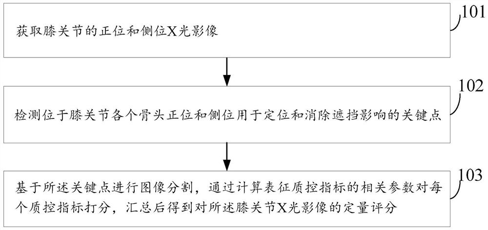
[0054] figure 1 A flow chart of a knee joint X-ray image quality control method according to an embodiment of the present invention, including the following steps:

[0055] Step 101, acquiring frontal and lateral X-ray images of the knee joint;

[0056] Step 102, detecting the key points located in the ortho and lateral positions of each bone of the knee joint for locating and eliminating the influence of occlusio...


PUM


Abstract

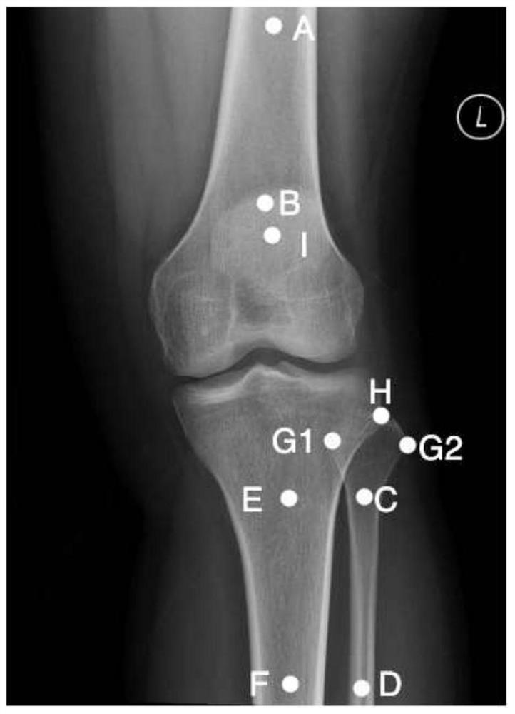
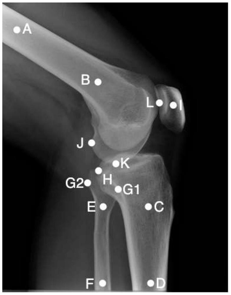
The invention provides a knee joint X-ray image quality control method and device. The method comprises the following steps: acquiring a normal position X-ray image and a lateral position X-ray image of a knee joint; detecting key points for positioning and eliminating shielding influence at the front position and the side position of each bone of the knee joint; and carrying out image segmentation based on the key points, scoring each quality control index by calculating related parameters representing the quality control indexes, and summarizing to obtain a quantitative score of the knee joint X-ray image. According to the method, a certain number of key points are marked at the normal position and the lateral position of each bone of the knee joint, so that positioning of each bone on the knee joint can be conveniently realized, and calculation of quality control indexes is simplified; due to the fact that segmentation, classification and other operations involved in quality control index calculation are carried out based on key points, most background areas can be removed, the memory occupation amount and the data calculation amount are reduced, and the running speed is increased; by setting a small number of redundant key points, the problem that quality control parameters are difficult to calculate due to shielding can be effectively solved.

Description

technical field [0001] The invention belongs to the technical field of medical imaging, and in particular relates to a knee joint X-ray image quality control method and device. Background technique [0002] Medical imaging technology (X-ray, CT, MRI, etc.) is the foundation of modern medical diagnosis technology. As a basic auxiliary diagnostic tool, it can help doctors to quickly locate the disease, so as to quickly make accurate and professional judgments. Digital Radiography (DR) is one of the most commonly used auxiliary methods by doctors due to its low price, less radiation to patients, and rapid imaging, especially for third-tier cities or some relatively backward cities. [0003] Due to the backwardness of local equipment, the small number of radiologists and the uneven professional level, the images taken are often not so perfect, and the quality of the images directly affects the final doctor's judgment and the determination of the later treatment plan. Traditiona...

Claims


Application Information

Patent Timeline
Patent Type & Authority Applications(China)
IPC IPC(8): G06T7/00G06T7/13G06T7/62
CPCG06T7/0012G06T7/13G06T7/62G06T2207/10116G06T2207/30008G06T2207/30168
Inventor 萧毅刘士远何福金刘小青俞益洲李一鸣乔昕王文文孙红标张茸李华
Owner HANGZHOU SHENRUI BOLIAN TECH CO LTD
Learn More