Imaging device and imaging system

A camera device and image technology, applied in the field of camera systems, can solve problems such as doctors are not easy

Pending Publication Date: 2022-06-21
阿伊里斯株式会社
View PDF0 Cites 0 Cited by
  • Summary
  • Abstract
  • Description
  • Claims
  • Application Information

AI Technical Summary

Problems solved by technology

However, the proper determination of influenza follicles requires intensive training in many cases, and it is by no means easy for ordinary doctors.

Method used


Image

  • Imaging device and imaging system
  • Imaging device and imaging system
  • Imaging device and imaging system

Examples


Embodiment Construction

[0028] Various embodiments of the present disclosure will be described with reference to the drawings. In addition, the same code|symbol is attached|subjected to the component common to drawing.

[0029]

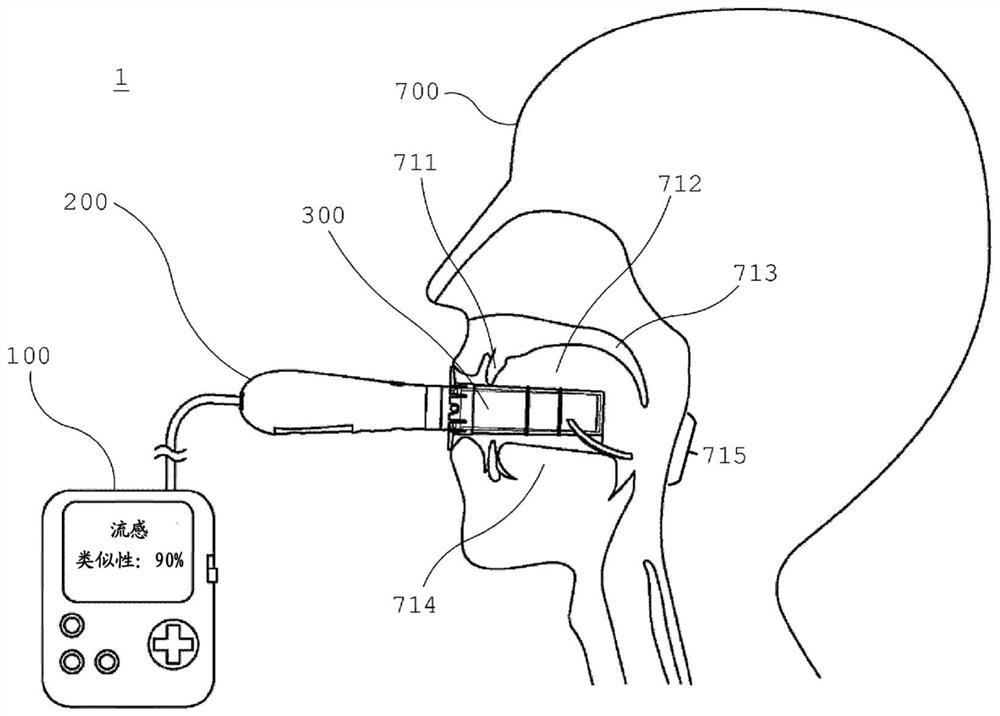
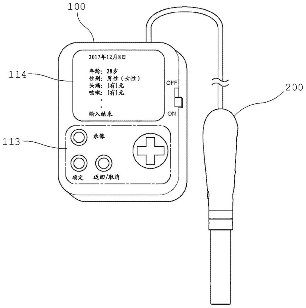
[0030] 1. Outline of camera system 1

[0031] The imaging system 1 of the present disclosure is mainly used for photographing the inside of a subject's oral cavity to obtain an image of the subject. In particular, the camera system 1 is used to photograph the periphery of the deep throat of the oral cavity, specifically, the throat. Therefore, the following mainly describes the case where the imaging system 1 of the present disclosure is used for imaging of the throat. However, the throat is an example of an imaging site, and of course, the imaging system 1 of the present disclosure can be applied to other sites as long as it is in the oral cavity.

[0032] As an example, the imaging system 1 of the present disclosure is used to image the throat of the oral cavity, and ...


PUM


Abstract

The present disclosure provides an imaging device and an imaging system suitable for diagnosis using an image of a subject obtained by imaging the inside of an oral cavity. Provided is an imaging device comprising: a main body which is provided with a base end and a distal end, and which is formed in a columnar shape having a predetermined length between the base end and the distal end in order to allow at least the distal end to be inserted into an oral cavity; one or a plurality of light sources disposed on the base end side or the tip end side of the main body, or the main body, and configured to irradiate light of a predetermined frequency band; a diffusion plate disposed on the base end side or the tip end side of the main body, or the main body, and configured so as to diffuse the light irradiated from the light source toward the oral cavity; and a camera which is disposed on the base end side or the tip end side of the main body, or the main body, and which captures an image of a subject on the basis of the reflected light diffused from the diffusion plate and reflected by the oral cavity.

Description

technical field [0001] The present disclosure relates to an imaging device and an imaging system for photographing the inside of the oral cavity of a subject person. Background technique [0002] Conventionally, it is known that a doctor observes changes in the oral cavity state of a subject, and diagnoses, for example, a viral cold. Non-Patent Document 1 reports that lymphoid follicles appearing in the deepest part of the throat located in the oral cavity have a pattern peculiar to influenza. Lymphoid follicles having this characteristic pattern are called influenza follicles, which are characteristic symptoms of influenza and appear about 2 hours after the onset of the disease. However, proper identification of influenza follicles requires intensive training in many cases, which is by no means easy for the general physician. [0003] prior art literature [0004] Non-patent literature [0005] Patent Document 1: Watanabe Miyamoto, "Investigation of the Significance and...

Claims


Application Information

Patent Timeline
IPC IPC(8): A61B1/267
CPCA61B1/24A61B1/000096A61B1/00188A61B1/00096A61B1/00039A61B1/00048A61B1/0638A61B1/000094A61B1/267A61B1/05A61B1/00066A61B1/00009A61B1/00016A61B1/00018A61B1/00131A61B1/0669
Inventor 安见卓志佐久间秋穂
Owner 阿伊里斯株式会社
Why Eureka
Try Eureka