Supercharge Your Innovation With Domain-Expert AI Agents!

Female pelvic floor ultrasonic model and preparation method thereof

A female pelvic floor and model technology, applied in the field of medical teaching models, can solve the problems of affecting the printing quality, insufficient mixing, and the inability to simulate the real situation of ultrasound examination, etc., and achieve the effect of increasing the printing quality

Inactive Publication Date: 2022-06-24
银川市第一人民医院
View PDF0 Cites 0 Cited by
  • Summary
  • Abstract
  • Description
  • Claims
  • Application Information

AI Technical Summary

Problems solved by technology

[0003] At present, only a few manufacturers can use PVC material to make female pelvis and pelvic floor muscle models, which can be used for daily learning by ultrasound operators and medical students, but the models can only be used to understand the organizational structure and cannot simulate the real situation during ultrasound examination , although ultrasonic diagnostic equipment is developing rapidly, but to a certain extent, the diagnostic accuracy rate still needs to rely on the operator's long-term accumulation and exploration to achieve high accuracy. For some rare lesions or structural damage, human factors have been greatly restricted. At present, 3D printers are used to prepare female pelvic floor ultrasound models. However, during the printing process of 3D printers, raw materials need to be mixed, but the mixing of one-way stirring can easily lead to insufficient mixing, which will eventually affect the printing quality.

Method used


Image

  • Female pelvic floor ultrasonic model and preparation method thereof
  • Female pelvic floor ultrasonic model and preparation method thereof
  • Female pelvic floor ultrasonic model and preparation method thereof

Examples


Embodiment Construction

[0028] The technical solutions in the embodiments of the present invention will be clearly and completely described below with reference to the accompanying drawings in the embodiments of the present invention. Obviously, the described embodiments are only a part of the embodiments of the present invention, but not all of the embodiments. Based on the embodiments of the present invention, all other embodiments obtained by those of ordinary skill in the art without creative efforts shall fall within the protection scope of the present invention.

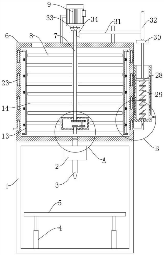
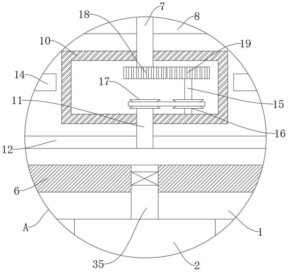
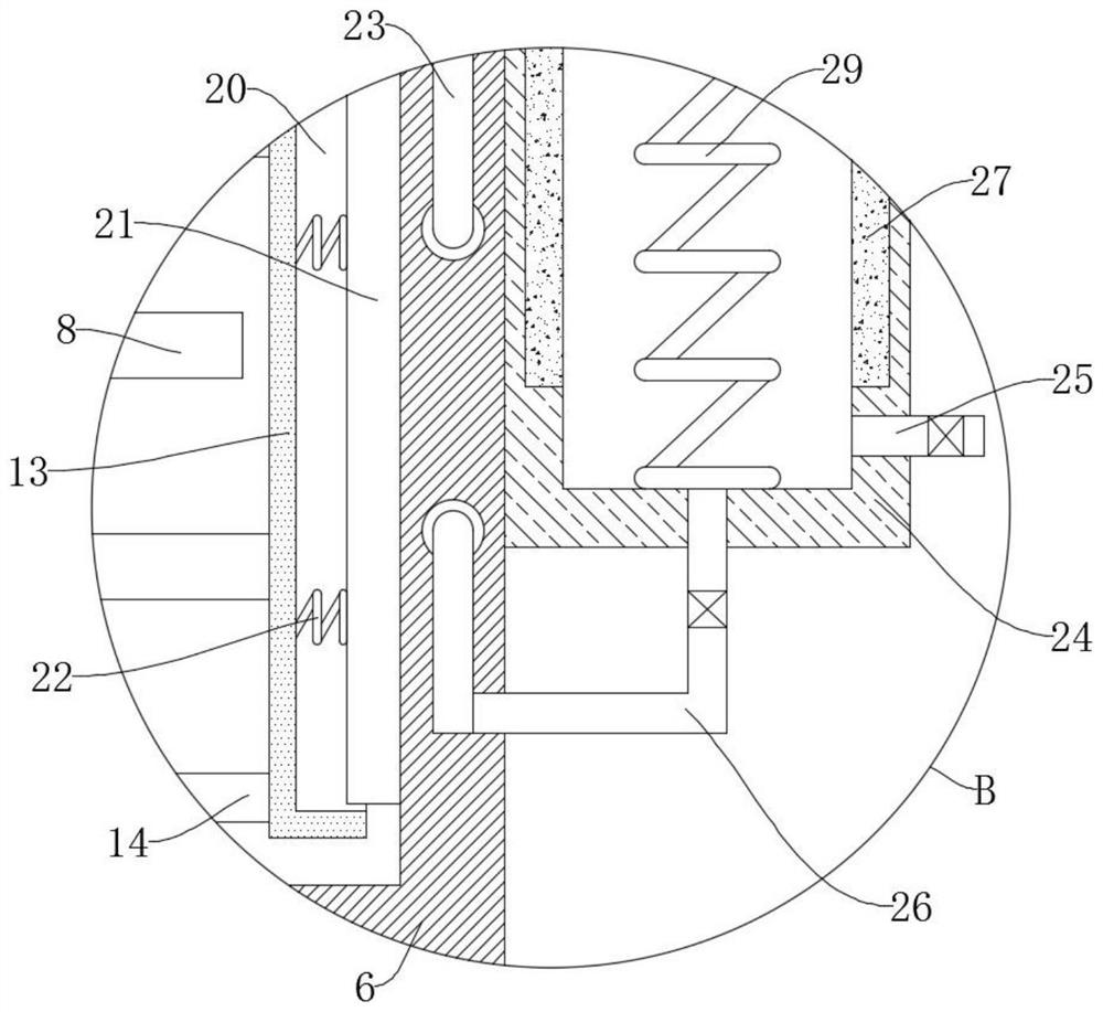
[0029] refer to Figure 1-4 , an ultrasonic model of female pelvic floor, including a 3D printer body 1, a control device 2 is installed on the top of the 3D printer body 1, a nozzle 3 is installed at the lower end of the control device 2, and the bottom of the 3D printer body 1 is connected with a bearing through a plurality of electric push rods 4. Plate 5, further, the control device 2 can control the nozzle 3 to move to print the ...


PUM


Abstract

The female pelvic floor ultrasonic model comprises a 3D printer body, the top of the 3D printer body is provided with control equipment, the lower end of the control equipment is provided with a spray head, the bottom of the 3D printer body is connected with a bearing plate through a plurality of electric push rods, and the upper end of the 3D printer body is fixedly connected with a mixing box; the invention further discloses a preparation method of the female pelvic floor ultrasonic model. The method comprises the steps that S1, a model printing material is added into a mixing box, then a pelvic floor model is printed out through a 3D printer body, obvious identification or color division is conducted on all parts, follow-up identification and operation are facilitated, and the model is placed in a container for use; according to the device, the rotating shaft can be driven to rotate in the opposite direction through rotation of the vertical rods, so that the rotating directions of the third rods and the first rods are opposite, a model printing material can be fully mixed and stirred, and the printing quality of a model is improved.

Description

technical field [0001] The invention relates to the technical field of medical teaching models, in particular to a female pelvic floor ultrasound model and a preparation method thereof. Background technique [0002] With the rapid development of medical ultrasound technology, ultrasonic diagnostic instruments such as Philips and other brand instruments are more functional, intelligent and concise, which greatly reduces the workload of operators and improves the efficiency and accuracy of inspections. Hospitals at all levels are gradually implementing Popularized and used, the female pelvic floor is composed of the levator ani muscle, the coccygeal muscle, and the fascia above and below the pelvic diaphragm and the fascia below the pelvic diaphragm covering the two muscles. Pelvic floor disorders, represented by pelvic organ prolapse, urinary incontinence, etc., plague the physical and mental health of about 30% of adult women. After a long-term follow-up investigation, the i...

Claims


Application Information

Patent Timeline
Patent Type & Authority Applications(China)
IPC IPC(8): G09B23/28B29C64/106B33Y10/00B33Y80/00
CPCG09B23/286B29C64/106B33Y10/00B33Y80/00
Inventor 马小萍鲍春旭刘东华虎桂玲高文涛段斌
Owner 银川市第一人民医院
Features
  • R&D
  • Intellectual Property
  • Life Sciences
  • Materials
  • Tech Scout
  • Unparalleled Data Quality
  • Higher Quality Content
  • 60% Fewer Hallucinations
Learn More