Closed cell culture system

A cell culture and closed technology, applied in tissue cell/virus culture devices, tissue culture, prostheses, etc.

Inactive Publication Date: 2006-12-27
新技术国际交流株式会社
View PDF2 Cites 2 Cited by
  • Summary
  • Abstract
  • Description
  • Claims
  • Application Information

AI Technical Summary

Problems solved by technology

In addition, it is difficult to collect tissues with complex three-dimensional structure at the damaged site, and infection is unavoidable during treatment, which has become an existing problem that has not yet been resolved

Method used


Image

  • Closed cell culture system
  • Closed cell culture system
  • Closed cell culture system

Examples


Embodiment 1

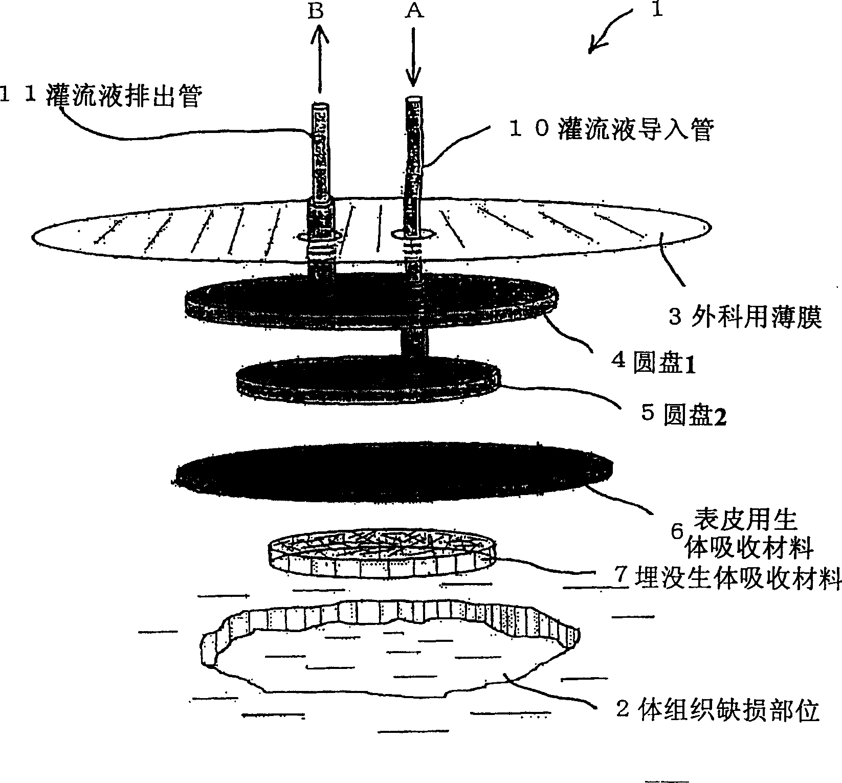
[0081] Embodiment 1: The treatment of the injured part after the operation of grafted skin

[0082] Use the blood fiber spray to quickly stop the bleeding from the wound surface after the skin picking operation, and disinfect the surrounding parts. Next, after filling the concave part with collagen particles, the open-cell collagen sheet with the hole opened in the center is made into a shape larger than the wound surface to cover the wound surface. The 2-layer adapter is also made into the same shape as the collagen sheet and stacked on top, covering the surgical film with holes only at the entrance and exit of the adapter to create a closed space for collagen particles and sheets. When there is pain at the skin picking site, circulate lidocaine as a local anesthetic and a plasma preparation containing 3 kinds of mixed antibiotics in a closed space, stop the local anesthetic and antibiotic solution after 3 hours, and switch to basic fiber Blast growth factor (bFGF) and perfu...

Embodiment 2

[0084] Example 2: Nerve reconstruction of severed nerves in lower extremity thigh

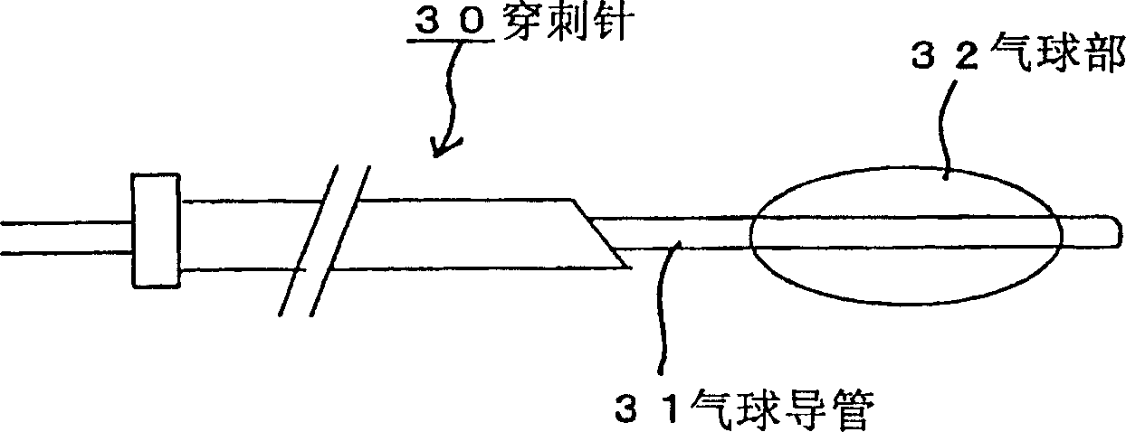
[0085] From the skin on the surface of the body, a softly bendable puncture needle is punctured to the nerves at both ends of the broken site of the nerve. The length of the balloon is selected to reach the length of the two broken ends of the nerve, the balloon catheter is inserted through the puncture needle to inflate the balloon, and a space is made between the broken ends of the nerve.

[0086] In this space, a catheter stored in the lumen is inserted into parallel collagen fiber bundles that form the basis of nerve growth. Furthermore, this catheter was exchanged with a small-diameter catheter for perfusion, the puncture needle was pulled out, and only the catheter for perfusion was left in place.

[0087] We add 10-8 moles of nerve growth factor to serum derived from patient's blood, and we start perfusion, and we switch circulatory circuit at the moment when internal air is exhausted. ...

Embodiment 3

[0089] Embodiment 3: the treatment of severe burn

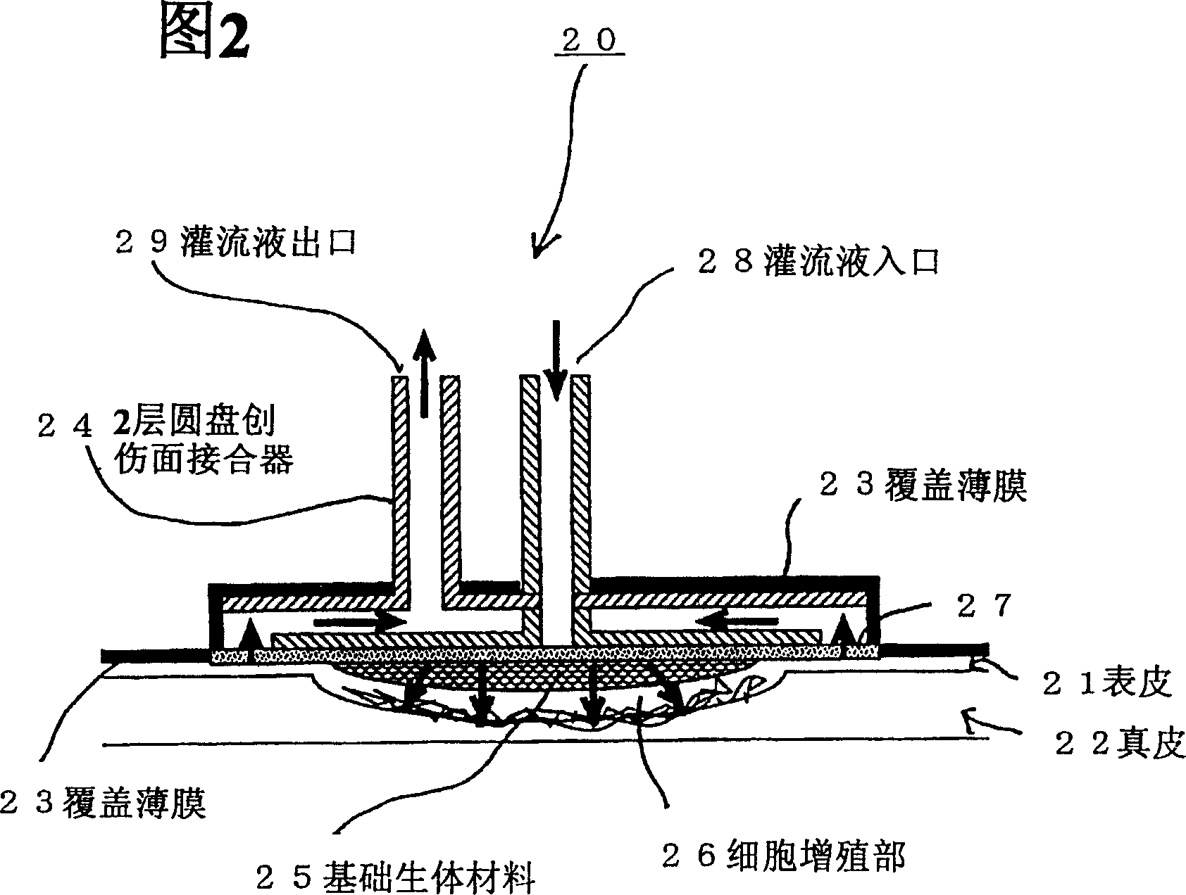
[0090] For severe burns on the abdomen, after removing skin and dead tissues, etc., cover the burn wound surface with a collagen fiber sheet, and then install a two-layer perfusion adapter on it. The wound surface was initially sterilized with an isotonic plasma-boost preparation containing povidone-iodine.

[0091] In order to inhibit the loss of body fluid from the patient's wound surface, an artificial plasma preparation with an osmotic pressure equal to that of the burn wound surface is perfused. Furthermore, in order to prevent infection, in addition to injection of antibiotics, if the patient complains of pain, lidocaine as a local anesthetic is injected into the perfusate for pain relief.

[0092] After the patient's condition stabilized, the administration of lidocaine and antibiotics was stopped, and cell growth factors such as fibroblast growth factor (bFGF), platelet-derived cell growth factor (pDGF), and epiderma...


PUM


Abstract

An object of the present invention is to provide a tissue cell culture system whereby a call tissue can be efficiently and quickly proliferated in vivo and the onset bacterial infection in an injured part can be avoided in the course of a treatment. More specifically, a closed cell culture system (1) characterized in that a defection (2) of a tissue on the body surface or inside the body is tightly sealed to form a closed environment free from the invasion of bacteria, etc. and then a solution appropriate for cell culture is circulated in the tissue defection thus sealed to thereby regenerate the defective tissue; and a method of administering a drug which comprises dissolving a remedy in the perfusion with the use of the above system and thus promoting the treatment of the defection.

Description

technical field [0001] The present invention relates to a system for inducing tissue regeneration at an injured site by proliferating cellular tissue at the injured site in vivo. Background technique [0002] In the past, when treating a damaged part of the body, after removing unnecessary substances such as foreign matter in the damaged part, disinfect it with a disinfectant, etc., rub the damaged part with a wound covering agent suitable for treatment, and achieve self-regeneration of the skin through cell proliferation . However, there are various insurmountable problems such as the time required for treatment in the self-regeneration of the skin, and the occurrence of infection at the wound site. [0003] In order to solve these problems, in recent years, as regenerative medicine, autologous tissue transplantation by culture has been adopted, that is, cells near the damaged site are collected and proliferated in vitro, and the target tissues and internal organs are reco...

Claims


Application Information

Patent Timeline
Patent Type & Authority Patents(China)
IPC IPC(8): A61L27/38C12N5/00A61L27/50C12M3/00A61L27/54A61L27/58A61L27/60A61L31/14
CPCA61F13/00068A61M25/10A61L2300/414A61F13/00063A61L27/54A61F2013/00536A61L27/3804A61L2300/406A61L27/60A61L31/148A61F2013/00927A61L27/58A61F2013/00174A61F2013/00221A61F2013/0054A61M37/00A61L27/3895A61L2300/402
Inventor 铃木茂树
Owner 新技术国际交流株式会社
Why Eureka
Try Eureka