Methods and compositions for molecular imaging
a molecular imaging and composition technology, applied in the field of compositions and methods for imaging targets, can solve the problems of not improving, the method currently does not exist for early detection of lung occult metastases, and the results are generally wors
- Summary
- Abstract
- Description
- Claims
- Application Information
AI Technical Summary
Benefits of technology
Problems solved by technology
Method used
Image
Examples
example 1
Imaging of Metastasized Human Breast Cancer Cells in Mouse Lung
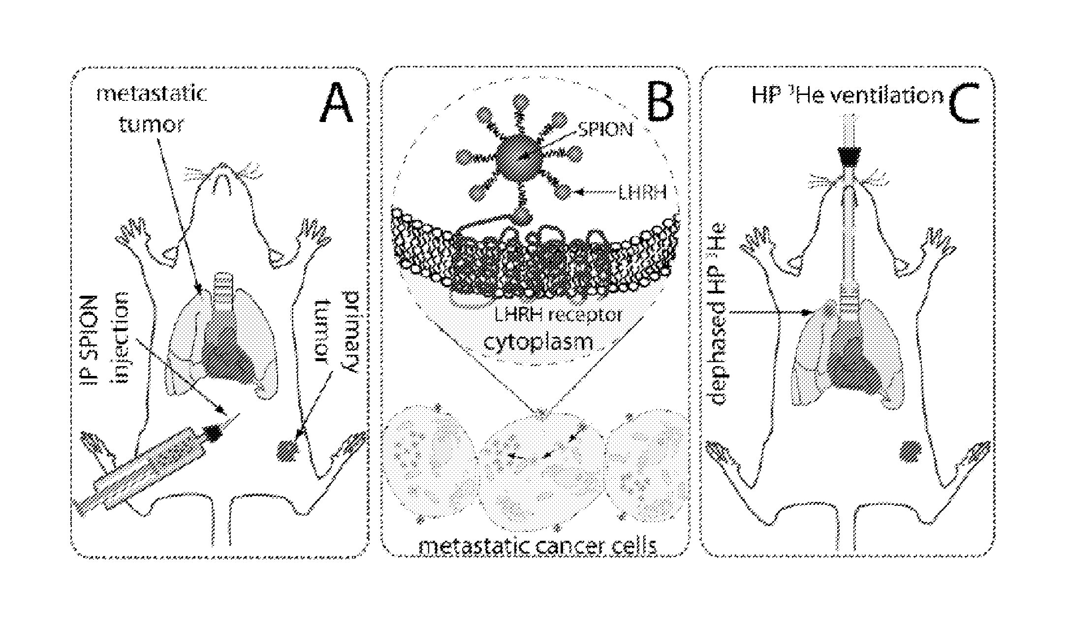
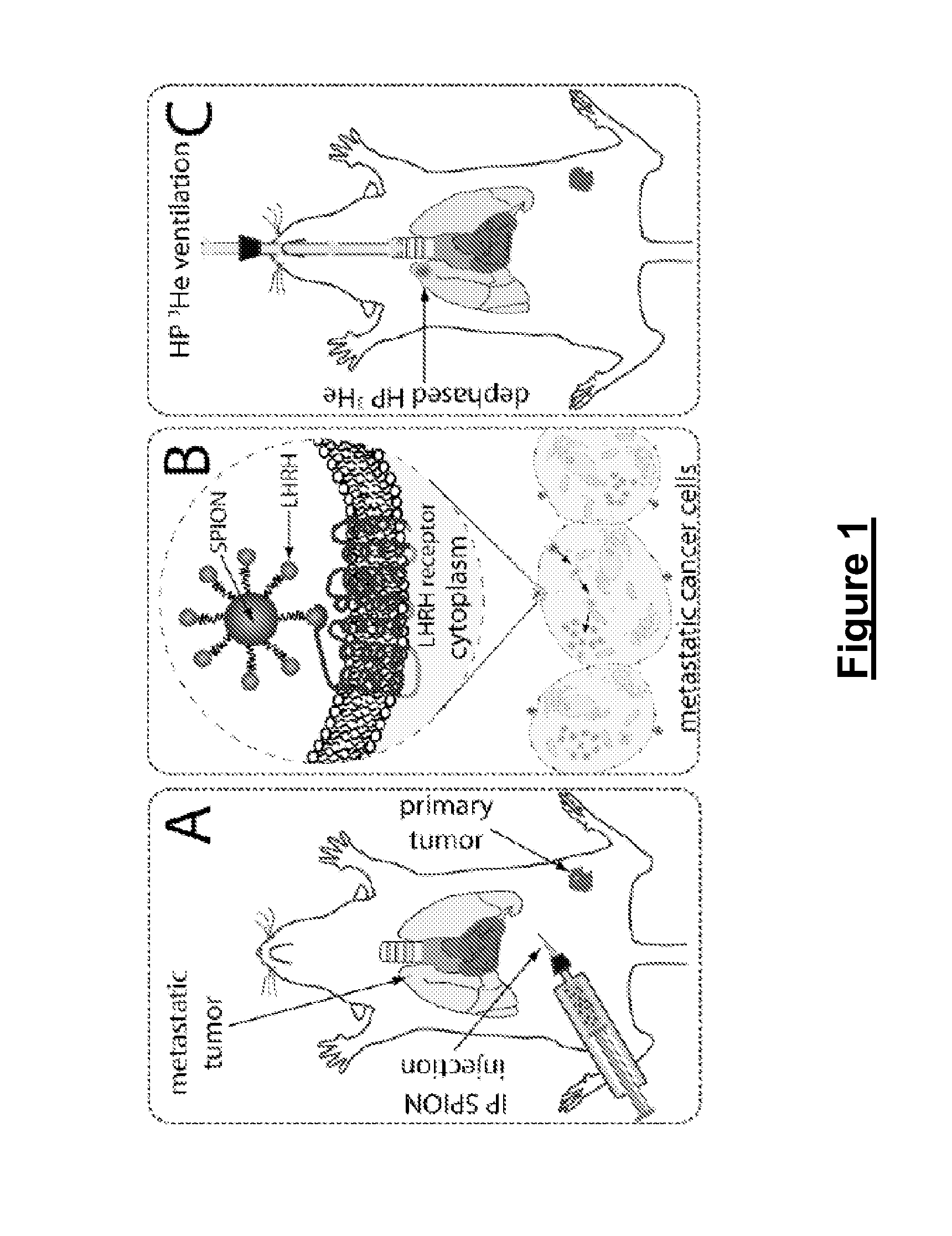
[0112]Female BALB / c nude mice at 9 weeks of age were inoculated with MDA-MB-435S human breast cancer cells. These cells metastasized to the lungs and expressed LHRH.
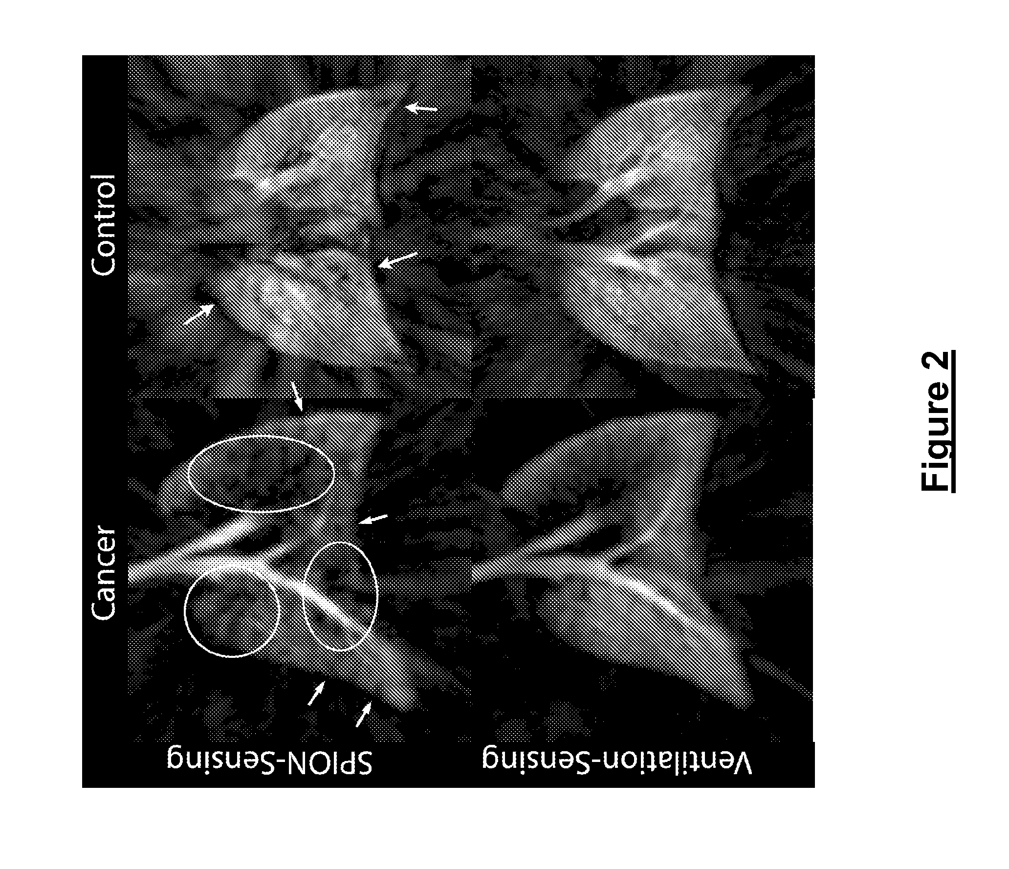
[0113]After about 60 days, and 24 hours before the imaging session, the mice received an intraperitoneal (i.p.) injection of 100 mg / kg LHRH-SPIONs (i.e., SPIONs to which LHRH peptides were conjugated). The mice were imaged using a 3D radial acquisition at a resolution of 156×156×1000 μm3. Images were acquired at TE=0.3 ms to show regional ventilation and TE=4 ms for T2* sensitivity. K-space was filled with 11,520 radial views acquired either 20 per breath with TR / TE=5 / 0.3 ms or 10 views per breath with TR / TE=10 / 4 ms. All images used BW=31.25 kHz and a fixed flip angle of 13° or 18°. After imaging, mice were sacrificed and lungs fixed for histology. Histology slides were stained with Prussian Blue to highlight iron so that a correlation could be made between d...
example 2
Imaging of Metastasized Human Prostate Cancer Cells in Mouse Lung
[0117]FIG. 4A shows an example of 3D gradient echo 3He images, at relatively long echo time (TE=4 ms), from a control animal that received a SPION injection but no tumor xenograft. FIG. 4B shows similar views of a mouse bearing a primary tumor produced by xenografted human prostate tumor cells. Whereas the control animal showed high signal intensity throughout the lungs, the tumor-bearing animal showed a pronounced signal loss in the right cranial lobe. Such signal voids were representative of all animals studied and were identified in 11 of the 12 tumor-bearing mice. In 10 of the 11 mice, the signal loss was located in the right cranial lobe, while in one mouse, the signal loss affected the entire right lung.
[0118]Comparison of 3He MRI against hematoxylin and eosin (H&E) histology (as shown in FIG. 5 for another prostate tumor model mouse) confirmed that 3He signal voids occurred at the site of metastatic tumors that ...
example 3
Positive Contrast SPION Detection
[0124]As disclosed herein, 3He imaging has successfully detected local SPION contrast through darkening of the image (negative contrast). There are also potential advantages to instead image SPIONs using positive contrast.
[0125]To this end, He MRI pulse sequences that allow the visualization of viable tumor tissue in mice with human breast or prostate cancer xenografts that have been targeted by LHRH-SPIONs have been developed. These pulse sequences made use of the magnetic field distortion created nearby the SPION particles to allow a background free detection of the targeted area.
[0126]An example of positive contrast SPION detection is presented in FIG. 7, which shows typical images acquired using a conventional spin-echo image, a T2 map, and a positive contrast image. The positive contrast particularly enhanced in the outer area of the primary tumor, later confirmed by histological examination, to be viable and loaded with LHRH-SPIONs.
[0127]A meth...
PUM
 Abstract
Description
Claims
Application Information
 - R&D
- Intellectual Property
- Life Sciences
- Materials
- Tech Scout
- Unparalleled Data Quality
- Higher Quality Content
- 60% Fewer Hallucinations
Browse by: Latest US Patents, China's latest patents, Technical Efficacy Thesaurus, Application Domain, Technology Topic, Popular Technical Reports.
© 2025 PatSnap. All rights reserved.Legal|Privacy policy|Modern Slavery Act Transparency Statement|Sitemap|About US| Contact US: help@patsnap.com


