
Automated image fusion/alignment system and method

An automatic overlapping and image technology, applied in image data processing, graphics and image conversion, measurement using nuclear magnetic resonance imaging system, etc.

Inactive Publication Date: 2007-04-18
COMPIZED MEDICAL SYST
View PDF2 Cites 3 Cited by
  • Summary
  • Abstract
  • Description
  • Claims
  • Application Information

AI Technical Summary

Problems solved by technology

Although these methods have shown some success, it is believed that the time to solution should be greatly improved without sacrificing accuracy

Method used


Image

  • Automated image fusion/alignment system and method
  • Automated image fusion/alignment system and method
  • Automated image fusion/alignment system and method

Examples


Embodiment Construction

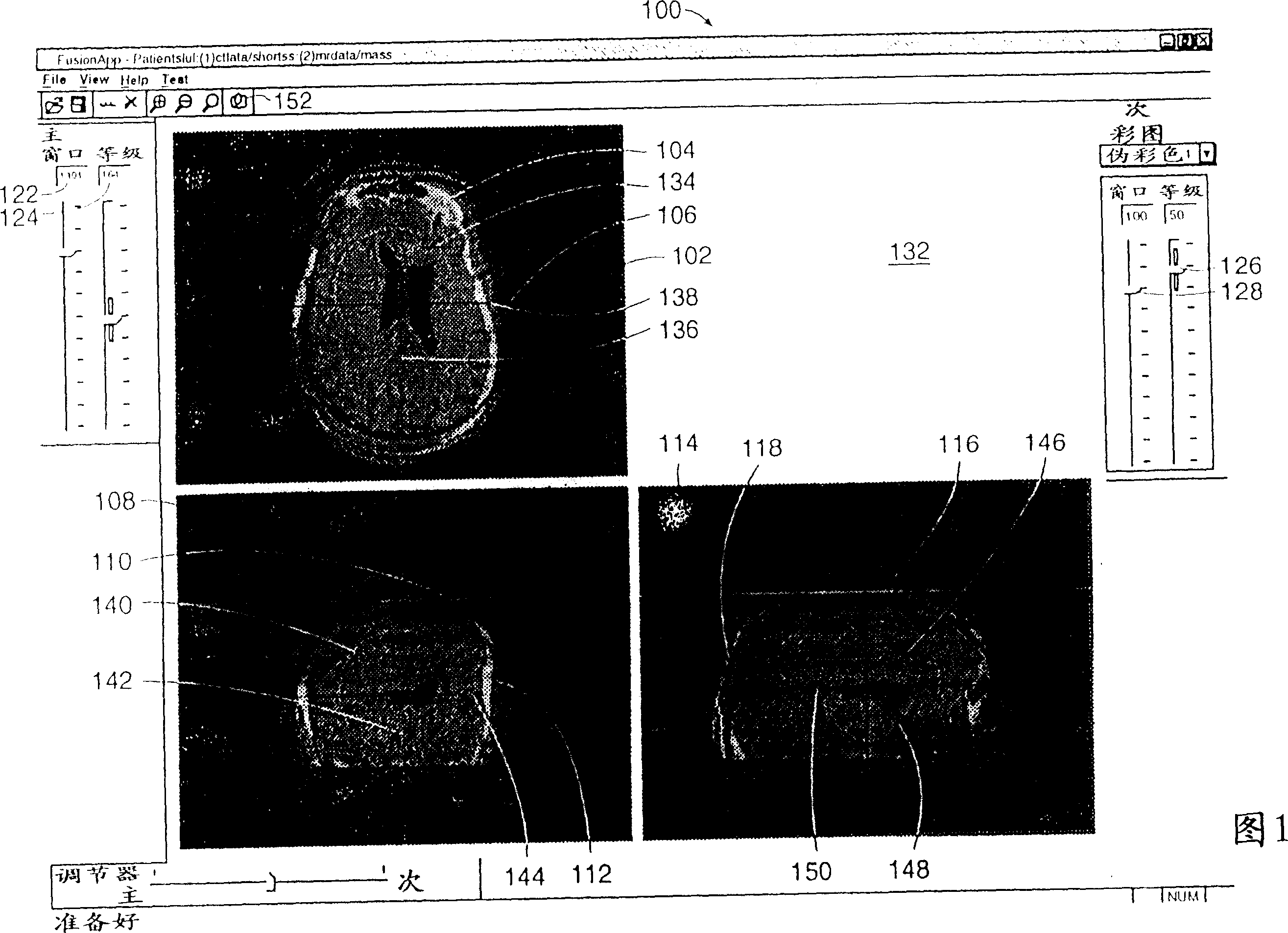
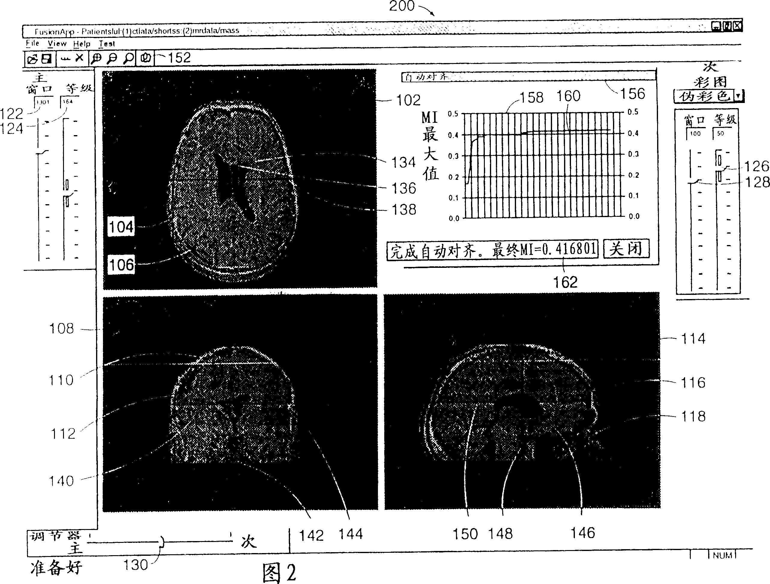
[0037] The present invention is a system and method for automatically registering three-dimensional ("3D") image data volumes. According to the system and method of the present invention, at least, but preferably two 3D image data volumes are simultaneously displayed on the GUI, and one 3D image data volume remains fixed and the other can be scaled, rotated and translated to align the coherent anatomy Features to achieve automatic coincidence. The systems and methods automatically display axial, sagittal and coronal views of the region of interest. These planar views preferably show the voxel intensity on a 2D plane within the data volume whose position is specified by the position of the linear cursor in the axial, coronal and sagittal windows of the GUI, e.g. "drag "The cursor within either window specifies a new location for the floor plan and updates the floor plan immediately without lag to the user. Under the second operation method, the cursor can be used to "drag" on...


PUM


Abstract

A system and method for autofusion of three-dimensional (3D) image data volumes is described. According to the system and method, registration is effected by simultaneously displaying on a GUI at least, but preferably, two 3D, image data volumes, and one of the 3D image data volumes is held fixed while the other may be scaled, rotated, and translated to align homologous anatomic features. The system and method will simultaneously display the axial, sagittal, and coronal plane views of a region of a region of interest. These plane views will be of data volumes that are designed and configured for a system user to ''drag'' or ''position'' one volume to a new position in one plane view which will also simultaneously update the data volume in the other two plane views with no apparent lag time. The system and method also includes automated alignment computation based on mutual information (MI) maximization. The system and method aligns 3D more efficiently and faster than prior image alignment methods.

Description

Technical field: [0001] The present invention relates to systems and methods for aligning volumes of image data. More specifically, the invention relates to aligning three-dimensional image data volumes. Background technique: [0002] In the medical field, it is often necessary to observe the internal organization of a handler with a large number of modalities. These modalities include x-ray imaging, magnetic resonance imaging ("MRI"), and computed tomography ("CT"). Other modalities exist such as functional magnetic resonance imaging ("fMRI"), single light emission computed tomography ("SPECT"), and positron emission computed tomography ("PET"), all of which contain images that describe living tissue. Physiological or metabolic information. [0003] It is known that the images produced by each modality have certain advantages and disadvantages. For example, radiographic ("X-ray") imaging has high spatial high-density resolution, shows bony anatomy in high detail, and is...

Claims


Application Information

Patent Timeline
Patent Type & Authority Patents(China)
IPC IPC(8): G06T3/00G01R33/54A61B5/00A61B5/055A61B6/03G06T19/00
CPCG06T3/0006G06T3/02
Inventor 莱恩·海巴德德莱克·G·莱恩
Owner COMPIZED MEDICAL SYST
Learn More