Acoustic-photo chromatography imaging method for multiple-element array electronic scanning biological tissue and apparatus thereof

An electronic scanning, biological tissue technology, applied in the directions of acoustic wave diagnosis, infrasound wave diagnosis, instruments for radiological diagnosis, etc. Differences and other issues, to achieve the effect of good adaptability, wide application range and fast speed

Inactive Publication Date: 2004-01-28
SOUTH CHINA NORMAL UNIVERSITY
View PDF0 Cites 15 Cited by
  • Summary
  • Abstract
  • Description
  • Claims
  • Application Information

AI Technical Summary

Problems solved by technology

Since the photoacoustic signal carries its rich internal characteristic information, it can be used to judge and identify its internal conditions. Compared with traditional ultrasonic detection or X-ray imaging methods, this method can distinguish the same acoustic impedance or X-ray absorption. For samples with different optical parameters, this is especially important for early diseased tissues, because the acoustic impedance and X-ray absorption characteristics of early diseased tissues (such as breast cancer) are not significantly different from those of surrounding normal tissues. The metabolism of the tissue is very different from that of the surrounding normal tissue; for example, the capillaries around the early cancerous tissue are much more abundant than the normal tissue, and the blood oxygen content is high, which causes it to have different optical properties from the surrounding normal tissue. If there is a large difference, photoacoustic signals can be used to image the difference in metabolic function between biological diseased tissue and normal tissue, so that diagnosis can be made and the accuracy of diagnosis can be improved; however, due to the complexity of biological tissues, photoacoustic signals in biological tissues Precise measurement of spores is a prerequisite for analysis, diagnosis and imaging of their characteristics
At present, when measuring with a detector, the acoustic sensor is basically placed on the end face of the sample to measure the photoacoustic signal transmitted from the sample; the signal obtained in this way is generally the signal generated by each sound point of the bulk acoustic field at the measurement point. Therefore, it is difficult to reconstruct the photoacoustic image in the measured body or judge the signal of the exact point inside it. It requires multi-point measurement and complex algorithm to process data, and the calculation amount is very large; and for the application of biological tissue, because it is A strong scatterer of light, the incident laser diverges quickly, and biological tissue is generally not an isotropic optical and acoustic body, so the sound field generated by the laser and the reflection, transmission and absorption of the sound field are very complicated, and it is difficult to obtain the The exact photoacoustic signal, so simplifying the measurement signal is one of the key issues for the application

Method used


Image

  • Acoustic-photo chromatography imaging method for multiple-element array electronic scanning biological tissue and apparatus thereof
  • Acoustic-photo chromatography imaging method for multiple-element array electronic scanning biological tissue and apparatus thereof
  • Acoustic-photo chromatography imaging method for multiple-element array electronic scanning biological tissue and apparatus thereof

Examples


Embodiment

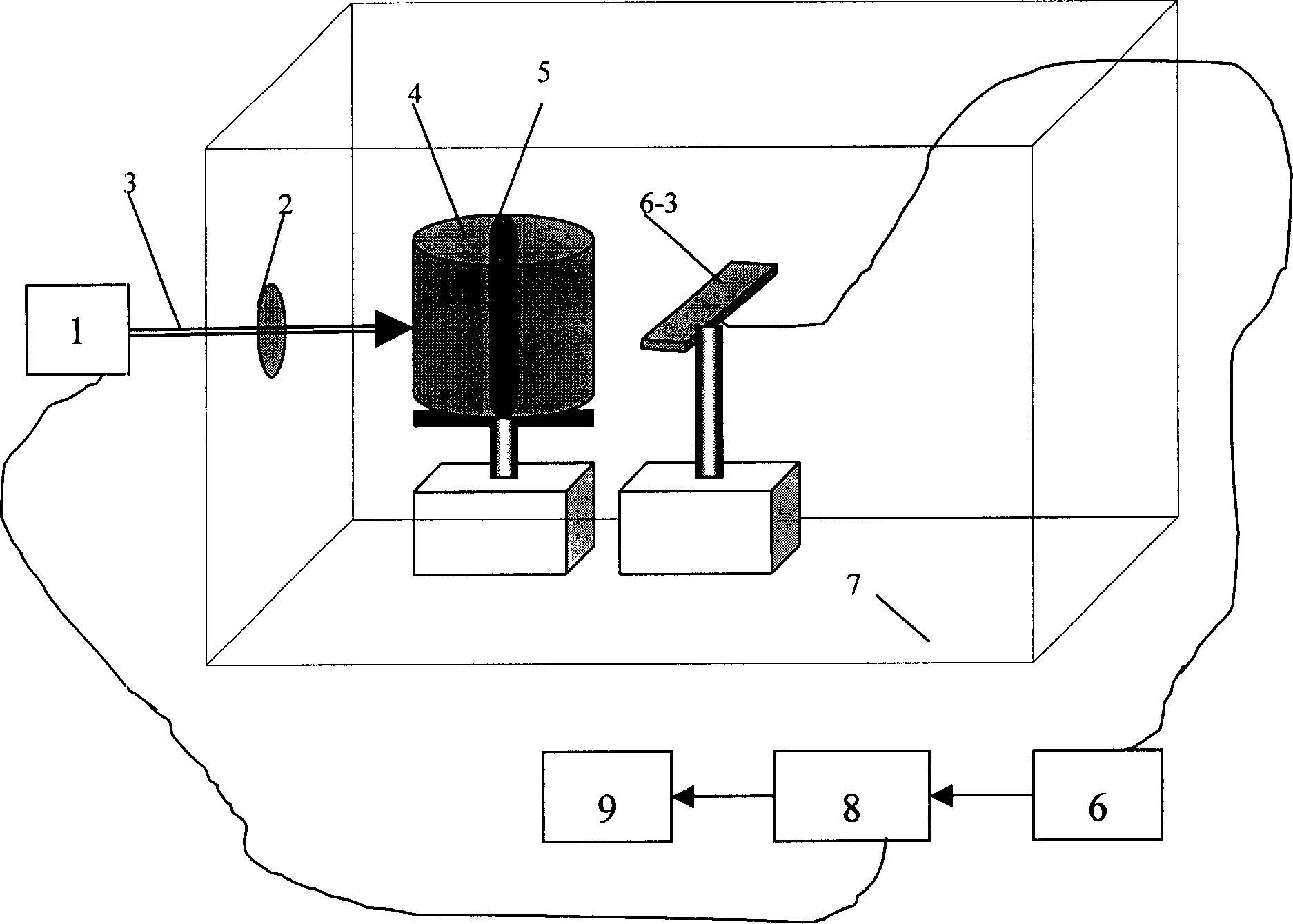
[0038] This embodiment mainly utilizes the 320-vibrator detector of the EUB-240 type ultrasonic diagnostic instrument, carries out appropriate modification to it, and constitutes a multi-element array electronic scanning detector, and forms a biological tissue with this multi-element array electronic scanning detector. A photoacoustic tomography device realizes photoacoustic tomography of multiple array electronic scanning biological tissues.

[0039]EUB-240 ultrasonic diagnostic instrument (hereinafter referred to as "B-ultrasound") is a multi-functional ultrasonic tomographic diagnostic device produced by Hitachi, Japan. It has strong measurement functions, multi-mode display functions and various images. Zoom function. Its 320 vibration element probe has the following characteristics: 1. Scanning method: linear array electronic scanning; 2. Nominal frequency: 5MHZ (3.5MHZ is also available); 3. Number of transducer vibration elements: 320 vibration elements / 80 groups ( 1 g...


PUM


Abstract

The invention provides a multielement array electronic scan bio-organization photoacostic layer separating imaging method and the equipment, including the steps: the pulse laser comes into the bio-tissue to produce photoacoustic signal which is collected and stored by computer; use multielement array electronic scan detector receives the photoacoustic signal, synchronously, and at the same time the computer collects and stores the photoacoustic signal; repeat collection until all the group elements are collected once; after finishing collecting, the computer makes filtering and integral processing on the optical-voice signal, and then performs layer separating imaging on the bio-organization by backward projecting algorithm. The equipment includes the laser, the multielement array electronic scan detector, the high-speed collection card and the computer. The equipment's automation level is high, convenient to operate and simpler to control and use.

Description

technical field [0001] The invention relates to photoacoustic tomography technology, in particular to a method and device for multiple array electronic scanning photoacoustic tomography of biological tissue. Background technique [0002] In recent years, research on the photoacoustic effect and photoacoustic tomography has received more and more attention. When a certain object is irradiated with light, its internal temperature will change due to its absorption of light, which will cause changes in the mechanical parameters of its local area; when a pulsed light source or modulated light source is used, the fluctuation of the local temperature inside the object will cause Its volume expands and contracts, thus radiating sound waves outward. This phenomenon is called photoacoustic field effect (referred to as photoacoustic effect). The photoacoustic effect is actually an energy conversion process. According to the heat conduction equation and wa...

Claims


Application Information

Patent Timeline
Patent Type & Authority Applications(China)
IPC IPC(8): A61B6/00A61B6/02A61B8/00
Inventor 邢达尹邦政王毅陈更生
Owner SOUTH CHINA NORMAL UNIVERSITY
Why Eureka
Try Eureka